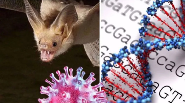
Kusetuskampanjan sisältö on ollut, että korona-virusryhmän ikiaikaisia, laajalle jakautuneita ominaisuuksia esitetään muka "mahdollisiksi syntyä vain laboratorio-olosuhteissa"...
Aivopierun lähettivät liikkeelle kaksi NATO:n-Bilderbergin-WEF:n infosoturia, (vale-)AIDS-rototetutkijat britti Angus Dalgleish ja norjalainen Birger Sørensen
Molemmat nämä herrat tekivät muuten "Halosen-Rissalan-ihmepelastuksen" vuonna 2014 Ukrainan sotatoimialueen yläpuolella (jonne ei missään tapauksessa olisi saa-nut matkustajakoneella lentää!) alasammutun Malaysian Airlinesin MH17-matkusta-jakoneen matkasta, jossa oli menossa EU-maiden delegaatio Melbournen AIDS-rokote-maailmankonferenssiin.
Onnettomuudessa kuoli 6 EU:n johtavaa (parhaiten rahoitettua, paitsi Dalgleish ja Sørensen ja Pekka Sillanaukee, €100 mlj.) AIDS-rokotetutkijaa mukaan lukien alan maailmanjärjestön ja Melbornen konferenssin hollantilainen puheenjohtaja Joep Lange ja lisäksi parikymmentä heidän assistenttiaan, sihteeriään ja perheenjäsentään. Rokotetta ei ole vieläkään - ja tuskin koskaan tuleekaan.
Koronavirus lähti todennäköisesti liikkeelle laboratoriosta, USA:n energiaministeriö uskoo nyt – Tiedustelujärjestöt ovat erimielisiä asiasta

Koronaviruksen omikronmuunnos.
***
Keskustelua:
Risto Koivula
23.05.2021 09:55:33
405355
Re: Pääministeri Marinkin myöntää koronapandemian olleen keinotekoinen
Lainaus: Teppolainen, 20.05.2021 15:57:34, 405340
https://www.iltalehti.fi/politiikka/a/32434a20-3629-40e7-9549-076d97a90f4b
Se vaan ei ole keinotekoinen, eikä Marinkaan tuossa sellaista väitä. Se ei myskään "johdu 1000-vuotisesta patriakaatista"...
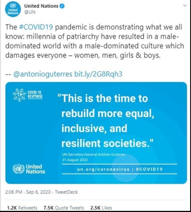
YK:n (EU-pölkky)pääsihteeri Guterrez:
"Koronavirus on 1000-vuotisen PATRIARKAATIN SYYTÄ!!!"
https://russian.rt.com/inotv/2020-09-08/Daily-Mail-gensek-OON-vozmutil
" Daily Mail: генсек ООН возмутил соцсети, связав пандемию с «тысячелетием патриархата»
Оригинал новости ИноТВ:
Daily Mail: генсек ООН возмутил соцсети, связав пандемию с «тысячелетием патриархата»
Оригинал новости ИноТВ:
В интернете распространяются призывы лишить ООН финансирования. Пово-дом для недовольства стало заявление главы международной организации о том, что пандемия коронавируса — «демонстрация патриархата», пишет Daily Mail.
Internetissä leviää vaatimus alentaa YK:n rahoitusta. Tyytymättömyyden lähde on kansainvälisen organisaation johtajan tiedotus siitä, että koronsaviruspandemia oa "patriarkaatin demonstraatio", kirjoittaa Daily Mail.
Facebook KIELTÄYTYI JULKASEMASTA TÄTÄ!!! "
Teppolainen
24.05.2021 11:41:18
405357
WSJ: Laboratoriossa joutui useita henkilöitä sairaalahoitoon COVID-19 taudin oireiden vuoksi ennen pandemian alkua
Yhdysvaltain mukaan useita henkilöitä sairastui Wuhanin viruslaboratoriossa mystisesti Covid19 taudin oireisiin ja joutui sairaalaan:
Myöskään Kiina ei suostu jutun mukaan avaamaan tietoja laboratoriossa tutkituista viruksista, jotta vaihtoehto laboratoriosta lähteneestä viruksesta voitaisiin poissulkea.
Se että tutkijoiden mukaan viruksen genomia ole tarkoituksellisesti muunneltu laboratoriossa, ei vielä tarkoita etteikö se olisi silti voinut olla lähtöisin laboratorio-olosuhteista.
...
Uusi tutkimus on julkaisemassa todisteita, joiden mukaan Covid olisi kiinalaisten tarkoituksella luoma virus, jonka jälkiä olisi peitelty muuntamalla virusta luonnollisemman oloiseksi:
https://www.foxnews.com/world/explosive-study-claims-to-prove-chinese-scientists-created-covid
Explosive study claims to prove Chinese scientists created COVID
British Professor Angus Dalgleish and Norwegian scientist Dr. Birger Sørensen wrote they’ve had primary evidence 'of retro-engineering in China'
A bombshell new study claims to have proof that Chinese scientists created COVID- 19 in a lab and then tried to reverse-engineer versions of the virus to make it look like it evolved naturally from bats.
British Professor Angus Dalgleish and Norwegian scientist Dr.Birger Sørensen wrote they’ve had primary evidence "of retro-engineering in China" since last year,but were ignored by academics and major medical journals, The Daily Mail reported Saturday, citing the soon-to-be-published study.
The study concludes: "the likelihood of it being the result of natural processes is very small." The virus is still killing 12,000 people a day around the world.
POMPEO SAYS WUHAN LAB WAS ENGAGED IN MILITARY ACTIVITY ALONGISDE CIVILIAN RESEARCH
Dalgleish is a London oncology professor known for breakthrough work on a vaccine for HIV. Sørensen is a virologist and chair of the pharmaceutical company Immunor, which developed a coronavirus vaccine candidate called Biovacc-19. Dalgleish also has a financial stake in that company.
It was during their COVID-19 vaccine research that the pair came across "unique fin-gerprints" indicating the virus didn’t come from nature, they said. The telltale clue: a rare finding in the COVID-carrying virus of a row of four amino acids, which give off a positive charge and bond to negative human cells.
INTEL COMMUNITY ‘AGGRESSIVELY’ INVESTIGATING COVID-19 ORIGIN
"The laws of physics mean that you cannot have four positively charged amino acids in a row," Dalgleish told the Daily Mail. "The only way you can get this is if you artificially manufacture it."
They also tracked published Chinese research, some done working with American universities, to show how the tools to create the virus were allegedly built. A good part of the work reviewed involved "gain of function" research, which involves mani-pulating natural viruses in a lab to make them more infectious, allowing scientists to study their potential effect on humans.
The U.S. put a moratorium on such research in 2014. But it’s impossible to know if $600,000 funding for medical research in China was used for gain of function research, Dr. Anthony Fauci told Congress last week. "
[RJK: Absolute bullshit!]
Viruksen varjolla kiinalaiset onnistuivat kiristämään totalitarismia omassa maassaan ja edistämään totalitarismia myös ulkomailla, jopa Suomessa, samalla kun heikentävät kilpailijamaitaan taloudellisesti.
On yhä selvempää, että Marinin hysteerinen koronastrategia on ollut täysin virheelli-nen - ja ulkomailta johdetulle Marinille tarpeeton totalitarismin lisääminen ei selvästi-kään ole tuottanut omantunnon tuskia. Marin ei ole edes yrittänyt edistää painostus-toimia Kiinaa vastaan, jotta viruksen alkuperä olisi saatu tyrehdytettyä. Edes sitä Marin ei ole tehokkaasti pyrkinyt estämään, että virusta tulisi Suomeen kolmansien maiden kautta. Sen sijaan Marin keskittyy rajoittamaan mielenosoitusoikeutta ja kokoontumisvapautta, ikään kuin se olisi ollut tehokkain mahdollinen keino viruksen leviämisen ehkäisemisessä.
***
Teppolainen
30.05.2021 14:38:32
405405
Pääministeri Marinin koronahysteria on yhä naurunalaisempi ajautuessaan kompukseen Kiinan kanssa
Lainaus: PeP, 30.05.2021 14:15:29, 405404
Lainaus: Teppolainen, 30.05.2021 11:34:17, 405400
On yhä selvempää, että Marinin hysteerinen koronastrategia on ollut täysin virheellinen - ja ulkomailta johdetulle Marinille tarpeeton totalitarismin lisääminen ei selvästikään ole tuottanut omantunnon tuskia.
On yhä selvempää,että Suomen hallitus oli valinnut aivan oikean linjan koronan vas- tatoimissa. Hallitus kuunteli asiantuntijoita syyllistymättä ylilyönteihin ja kykeni nou-dattamaan tehokasta strategiaa koronan torjunnassa. Joistain virheistä huolimatta sekä sairastuvuus, että koronakuolleisuus ovat olleet Suomessa Euroopan alhaisim-pia. Samaan aikaan talouden notkahdus on ollut useita Euroopan maita pienempi. Hallitukselle voi antaa koronan hoitamisessa hyvän arvosanan kaikenkarvaisten salaliittoteoreetikkojen huuhailusta huolimatta.
Teppolainen: "Nämä mainitsemasi asiat tapahtuivat totalitaristi Marinin hallituksesta huolimatta, ei hysteerisen koronastrategian ansiosta.
On yhä selvempää, että Marinin kaltaisia hyypiöitä ajavat täysin muut asiat kuin ih-misten hyvinvointi, kokoontumisenvapaus, sananvapaus ja muut yleisesti arvostetut asiat."
Risto Koivula
30.05.2021 15:30:15
405406
Tavallisia koronavirusryhmän ominaisuuksia esitetään "mahdottomina muualla paitsi laboratoriossa"
Lainaus: Teppolainen, 30.05.2021 11:34:17, 405400
https://www.foxnews.com/world/explosive-study-claims-to-prove-chinese-scientists-created-covid
Viruksen varjolla kiinalaiset onnistuivat kiristämään totalitarismia omassa maassaan ja edistämään totalitarismia myös ulkomailla, jopa Suomessa, samalla kun heikentävät kilpailijamaitaan taloudellisesti.
On yhä selvempää, että Marinin hysteerinen koronastrategia on ollut täysin virheelli-nen - ja ulkomailta johdetulle Marinille tarpeeton totalitarismin lisääminen ei selvästi-kään ole tuottanut omantunnon tuskia. Marin ei ole edes yrittänyt edistää painostus-toimia Kiinaa vastaan, jotta viruksen alkuperä olisi saatu tyrehdytettyä. Edes sitä Marin ei ole tehokkaasti pyrkinyt estämään, että virusta tulisi Suomeen kolmansien maiden kautta. Sen sijaan Marin keskittyy rajoittamaan mielenosoitusoikeutta ja kokoontumisvapautta, ikään kuin se olisi ollut tehokkain mahdollinen keino viruksen leviämisen ehkäisemisessä. "
Nuo tiedot ovat humpuukia, samoin nämä:
https://nypost.com/2021/05/29/explosive-study-claims-to-prove-chinese-scientists-created-covid/
Explosive study claims to prove Chinese scientists created COVID
By Eileen AJ Connelly
May 29, 2021
A bombshell new study claims to have proof that Chinese scientists created COVID- 19 in a lab and then tried to reverse-engineer versions of the virus to make it look like it evolved naturally from bats.
British professor Angus Dalgleish and Norwegian scientist Dr. Birger Sørensen wrote they’ve had primary evidence “of retro-engineering in China” since last year, but were ignored by academics and major medical journals, the Daily Mail reported Saturday, citing the soon-to-be-published study.
The study concludes: “the likelihood of it being the result of natural processes is very small.” The virus is still killing 12,000 people a day around the world.
Dalgleish is a London oncology professor known for breakthrough work on a vaccine for HIV. Sørensen is a virologist and chair of a pharmaceutical company, Immunor, which developed a coronavirus vaccine candidate called Biovacc-19. Dalgleish also has a financial stake in that company.
It was during their COVID-19 vaccine research that the pair came across “unique fin-gerprints” indicating the virus didn’t come from nature, they said. The telltale clue: a rare finding in the COVID-carrying virus of a row of four amino acids, which give off a positive charge and bond to negative human cells.
Some scientists believe the virus could have emanated from the Wuhan Institute of Virology in China.AFP via Getty Images
“The laws of physics mean that you cannot have four positively charged amino acids in a row,” Dalgleish told the Daily Mail. “The only way you can get this is if you artificially manufacture it.”
They also tracked published Chinese research, some done working with American universities,to show how the tools to create the virus were allegedly built.A good part of the work reviewed involved “gain of function” research, which involves manipula-ting natural viruses in a lab to make them more infectious,allowing scientists to study their potential effect on humans.
The US put a moratorium on such research in 2014. But it’s impossible to know if $600,000 in funding for medical research in China was used for gain of function research, Dr. Anthony Fauci told Congress last week.

British professor Angus Dalgleish (pictured) and Norwegian scientist Dr. Birger Sørensen claim to have proof COVID was created in a lab.
Chris Radburn/PA Wire/ZUMAPRESS.com
“A natural virus pandemic would be expected to mutate gradually and become more infectious but less pathogenic, which is what many expected with the COVID-19 pandemic but which does not appear to have happened,” the scientists wrote.
The scientists could not immediately be reached by The Post for comment.
News of the study comes amid renewed interest in COVID-19’s origins, which had long been proclaimed to have made the jump from bats to humans at a wet market in China. "
Oikeat tiedot löytyvät täältä:
https://www.sciencedirect.com/science/article/pii/S1873506120304165
" Stem Cell Research
Volume 50, January 2021, 102115
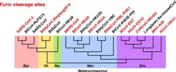
Furin cleavage sites naturally occur in coronaviruses
Author links open overlay panel Yiran Wu a, Suwen Zhao b:
https://doi.org/10.1016/j.scr.2020.102115
Under a Creative Commons license
Highlights
• Phylogenetic tree of spike proteins reveals major groups of coronaviruses.
• Furin cleavage sites at spike S1/S2 are common in coronaviruses.
• Furin cleavage sites at spike S1/S2 naturally occurred independently for multiple times in coronaviruses.
Abstract
The spike protein is a focused target of COVID-19, a pandemic caused by SARS-CoV-2. A 12-nt insertion at S1/S2 in the spike coding sequence yields a furin clea-vage site, which raised controversy views on origin of the virus. Here we analyzed the phylogenetic relationships of coronavirus spike proteins and mapped furin recog-nition motif on the tree. Furin cleavage sites occurred independently for multiple times in the evolution of the coronavirus family, supporting the natural occurring hypothesis of SARS-CoV-2.
1. Introduction
The coronavirus disease 2019 (COVID-19) pandemic, caused by coronavirus SARS-CoV-2,is still rapidly spreading. Scientists all around the world are studying the infec- tion process of the virus and looking for therapeutic solutions to prevent and cure the disease. Spike protein is one of most important targets in COVID-19 research, not only in mechanism study but also in vaccine development and therapeutic antibody design (Premkumar et al., 2020, Abraham, 2020). The spike protein forms a homo-trimer (Fig. 1A) and protrudes outside the membrane of the virion (Li, 2016, Wrapp et al., 2020, Walls et al., 2020). It binds to angiotensin-converting enzyme 2 (ACE2) on human cell surface, undergoes conformational changes, and mediates the fusion of the virion to human cells (Li et al., 2003, Yan et al., 2020, Lan et al., 2020, Wang et al., 2020, Benton et al., 2020).
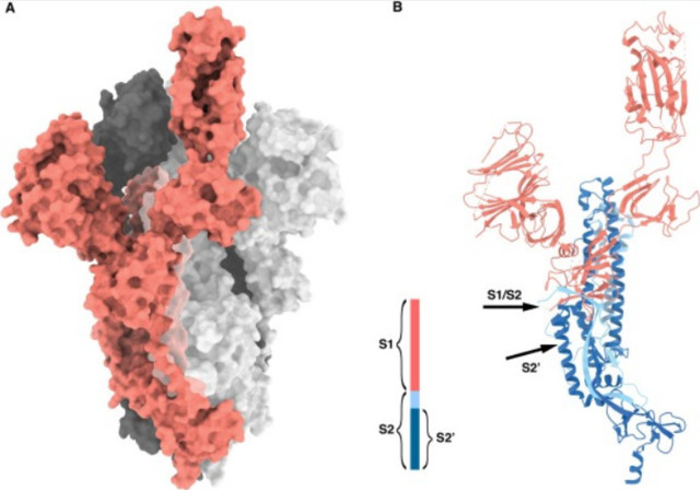
Fig. 1. Structure of SARS-CoV-2 spike protein (PDB ID: 6VYB (Walls et al., 2020). A) Spike homotrimer in open conformation. B) Cleavage sites on spike protein (marked by arrows). Red cartoon, S1; light/dark blue cartoon, S2; dark blue cartoon, S2′). (For interpretation of the references to colour in this figure legend, the reader is referred to the web version of this article.)
The spike protein consists of two subunits: the N-terminal S1 subunit, containing the receptor binding domain that interacts with ACE2 and induces conformational change; while the C-terminal S2 subunit,containing the fusion peptide that is respon- sible for fusing the membranes of virion and human cells (Li, 2016). To function pro-perly, the spike protein needs to be cleaved by host proteases to separate the two subunits (Simmons et al., 2013). Study of SARS-CoV (caused the severe acute res-piratory syndrome in 2002-2003 and belonged to the same species as SARS-CoV-2) showed that cleavage at S1/S2 enhances fusogenicity of spike protein (Bosch et al., 2003). Another cleavage site producing S2′ (fusion peptide and the C-terminal region of S2) was also identified (Fig.1B) (Belouzard et al.,2009).The S2′ site is not far from the S1/S2 site, and cleavage either or both them can yield the separation of two sub-units of spike.For coronaviruses,multiple proteases were reported responsible for the cleavage, including TMPRSS2 (Hoffmann et al.,2020, Glowacka et al.,2011); cathep- sin CTSL (Bosch et al., 2008, Ou et al., 2020), and trypsin (Belouzard et al., 2009, Bertram et al., 2011).
The spike protein of the Middle East respiratory syndrome coronavirus (MERS-CoV) was discovered to be more effectively cleaved by another protease, furin (Millet and Whittaker, 2014). In MERS-CoV,both S1/S2 and S2′ sites have sequence RXXR, the furin recognition motif. In human body, furin is ubiquitously expressed (Braun and Sauter, 2019). This may explain the highly lethal nature and high rate of multiple system failure caused by MERS (Millet and Whittaker, 2014).
Surprisingly, SARS-CoV-2 has the furin recognition motif at S1/S2, causing by a 12-nucleotide insertion not presented even in its closest relatives (Walls et al., 2020, Coutard et al., 2020). This stimulates a conspiracy that this furin site can only be ma-nual work, thus SARS-CoV-2 must be created in a laboratory. Here, we analyzed the sequences of coronaviruses and found furin sites occurred independently for mul-tiple times during evolution. This exhibits natural occurrence of furin cleavage site in SARS-CoV-2 spike protein is highly possible. Thus, the insertion of furin cleavage site into SARS-CoV-2 spike protein is not necessarily a result of manual work.
2. Results
2.1. Phylogenetic tree of spike protein sequences reveals major groups of coronaviruses
Coronaviruses are members of the subfamily Orthocoronavinae (also named Coro-navinae) in the family Coronavidae. There are four genera: Alphacoronavirus, Beta-coronavirus, Gammacoronavirus, and Deltacoronavirus. The first two genera only in-fect mammals, while the last two primarily infect birds but also mammals (Cui et al., 2019). We collected sequences of coronavirus spike proteins from the InterPro data-base (Mitchell et al., 2019) and performed phylogenetic analysis. The phylogenetic tree (Fig. 2A) generally matches reported relationship of coronaviruses (Cui et al., 2019): three genera (Betacoronavirus; Gammacoronavirus, and Deltacoronavirus) each forms a single clade,while only Alphacoronavirus has a small group left outside (see Fig. 5 for representative sequences). The left-out Alphacoronavirus group con-tains Rhinolophus bat coronavirus HKU2 that was reported to be closely related to two typical Alphacoronavirus, human coronavirus NL63 and human coronavirus 229E, in a phylogenetic analysis based on the RNA-dependent RNA polymerase coding region (Cui et al., 2019). We aligned the whole genomes of the three viruses and found Rhinolophus bat coronavirus HKU2 is quite unique in its spike protein-coding region (Fig. S1). Such result explains why this clade was separated from other Alphacoronavirus in our phylogenetic analysis.
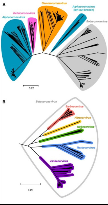
Fig. 2. Phylogenetic tree of coronavirus spike protein sequences. A) Noting genera of coronavirus. B) Subtree of Betacoronavirus, noting subgenera.
Furthermore, the five subgenera of Betacoronavirus were nicely displayed as mono-phyletic (Fig. 2B): Sarbecovirus (e.g. SARS-CoV-2 and SARS-CoV), Merbecovirus (e.g. MERS-CoV), Embecovirus (e.g. human coronavirus OC43 and human corona-virus HKU1, both causing common cold), and two small subgenera Hibecovirus and Nobecovirus. Hibecovirus and Nobecovirus are only discovered in bats so far, and our phylogenetic analysis shows they are related to Sarbecovirus.
2.2. Furin cleavage site at spike S1/S2 also occurs in a virus close to Sarbecovirus
We mapped the furin recognition motif RXXR at both S1/S2 and S2′ positions in phy-logenetic trees of spike protein sequences (Fig. 3, Fig. 4, Fig. 5 and S2, S3). In the Sarbecovirus + Hibecovirus + Nobecovirus clade (Fig. 3), furin cleavage sites at either position occur only in limited ranges. Strains of SARS-CoV-2 (we also added sequences from the GISAID database) have furin cleavage sites at spike S1/S2.
Moreover, SARS-CoV-2 is the only virus in subgenus Sarbecovirus having this feature, while even its closest relatives, bat coronavirus RaTG13 (sequence identity 97.7%) and pangolin coronaviruses (92.9%–90.7%), do not have furin site. However, in Hibecovirus, the sister clade of Sarbecovirus, a Hipposideros bat coronavirus col-lected in 2013 at Zhejiang Province in China has furin site at S1/S2. Interestingly, the other member in Hibecovirus lacks such site, similar to the situation of SARS-CoV-2 and its close relatives. The SARS-CoV-2 strains and Hipposideros bat coronavirus (Zhejiang 2013) are not sister groups, in agreement with the distinct sequence pat-terns of their furin cleavage sites at spike S1/S2 (Fig. 6A). Besides, two strains of bat coronavirus HKU9, belonging to Nobecovirus, are the only members in the Sarbeco-virus + Hibecovirus + Nobecovirus clade having furin cleavage site at spike S2′. ..."
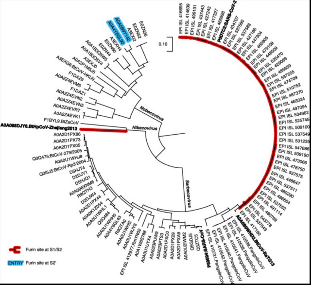
Fig. 3. Mapping of furin recognition motif on phylogenetic tree of spike protein sequences, the Sarbecovirus + Hibecovirus + Nobecovirus clade. Sequences in the SARS-CoV-2 clade were clustered with 95% identity threshold; in the SARS-CoV-2 clade were clustered with 99% identity threshold.
Kuvasta ilmenee, että kyseinen "Furin cleavage sites -naula" tai "-piikki" esiintyy riippumattomasti coronavirusten evoluutiossa ja lisäksi ERI PAIKOISSA eri viruksilla. Kaikilla noilla viruksilla ei ole tekemistä ihmisen kanssa, joskin tuo ryhmä vaikuttaa olevan ihmiselle vaarallinen tarttuvuuden edistäjänä.
... Furin cleavage sites naturally occur in coronaviruses
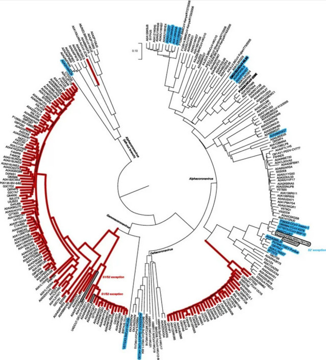
Fig. 5. Mapping of furin recognition motif on phylogenetic tree of spike protein sequences, the Alphacoronavirus + Gammacoronavirus + Deltacoronavirus clade. Sequences clustered with 95% identity threshold.
Vastaava kartoitus Alphacoronavirus + Gammacoronavirus + Deltacoronavirus -tyypeillä.
" ... 2.2. Furin cleavage site at spike S1/S2 also occurs in a virus close to Sarbecovirus
We mapped the furin recognition motif RXXR at both S1/S2 and S2′ positions in phylogenetic trees of spike protein sequences (Fig. 3, Fig. 4, Fig. 5 and S2, S3). In the Sarbecovirus + Hibecovirus + Nobecovirus clade (Fig. 3), furin cleavage sites at either position occur only in limited ranges. Strains of SARS-CoV-2 (we also added sequences from the GISAID database) have furin cleavage sites at spike S1/S2.
Moreover, SARS-CoV-2 is the only virus in subgenus Sarbecovirus having this feature, while even its closest relatives, bat coronavirus RaTG13 (sequence identity 97.7%) and pangolin coronaviruses (92.9%–90.7%), do not have furin site. However, in Hibecovirus, the sister clade of Sarbecovirus, a Hipposideros bat coronavirus col-lected in 2013 at Zhejiang Province in China has furin site at S1/S2. Interestingly, the other member in Hibecovirus lacks such site, similar to the situation of SARS-CoV-2 and its close relatives. The SARS-CoV-2 strains and Hipposideros bat coronavirus (Zhejiang 2013) are not sister groups, in agreement with the distinct sequence pat-terns of their furin cleavage sites at spike S1/S2 (Fig. 6A). Besides, two strains of bat coronavirus HKU9, belonging to Nobecovirus, are the only members in the Sarbeco-virus + Hibecovirus + Nobecovirus clade having furin cleavage site at spike S2′.
2.3. Furin cleavage sites are common in Betacoronavirus
Our mapping results showed that the furin recognition motif is more common in Merbecovirus and Embecovirus (Figs. 4 and S2, S3). In Merbecovirus, furin sites at spike S1/S2 occur in three clades: MERS-CoV strains, the bat coronavirus HKU5 strains, and coronavirus Neoromicia/PML-PHE1/RSA/2011 with its relatives (Figs. 4A and S2). Besides, MERS-CoV and bat coronavirus HKU5 are the only clades in Merbecovirus having furin cleavage site at S2′.
In Embecovirus, furin recognition motif at spike S1/S2 is universal: All strains but a few exceptions have furin cleavage sites at spike S1/S2 (Figs. 4B and S3). Interes-tingly, the Longquan Aa mouse coronavirus (Wang et al., 2015) loses this furin site, while its close relatives (e.g. China Rattus coronavirus HKU24, sequence identity 96.0%) maintains the furin cleavage site. This provides an example of naturally oc-curred sequence variation at spike S1/S2 among closely related coronaviruses. Be-sides, for spike S2′, only several single strains have furin recognition motif (Fig. S3).
2.4. Furin cleavage sites also occur in other genera of coronavirus
Our mapping results showed furin cleavage sites are widely present in the whole co-ronavirus family (Fig. 5). For spike S1/S2, furin recognition motif is universal in Gam-macoronavirus, and also occurs in two clades of Alphacorovanvirus: feline coronavi-rus and relatives, and Chevrier's field mouse coronavirus. For spike S2′, furin recog-nition motif occurs in several independent clades, covering all the three genera. Notably, in the two human coronaviruses in Alphacoronavirus causing common cold, HCoV NL63 has furin cleavage site at spike S2′, while the HCoV 229E (protein sequence identity 63.8%) lacks such feature.
...
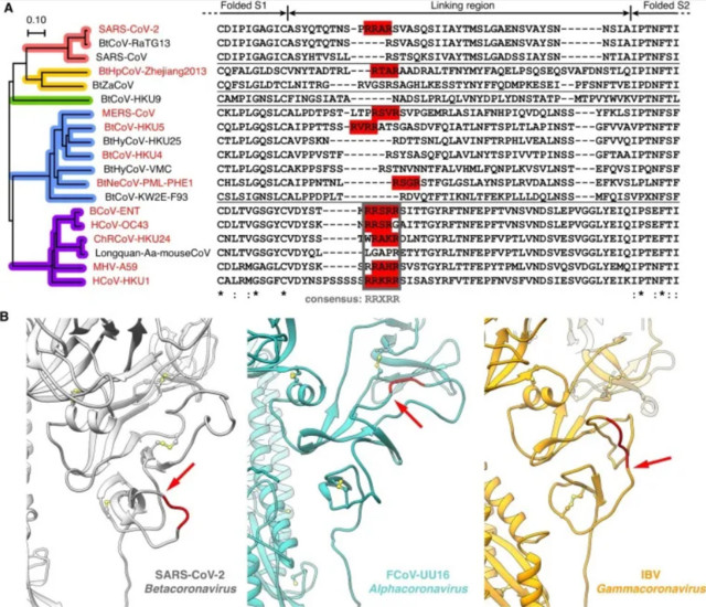
Fig. 6. Positions of furin cleavage sites at the linking region of S1 and S2. A) Multiple sequence alignment of representative Betacoronavirus spike protein S1/S2 region, with furin recognition motifs highlighted (red colorboxes in sequence alignment). Phylogenetic tree of spike protein se-quences is colored to indicate subgenera (coloring scheme the same as in Fig. 2B). B) Positions of furin cleavage sites in different coronavirus genera (red cartoon, furin recognition motif; red arrow, cleavage site); structures: SARS-CoV-2, PDB ID 6VYB (Walls et al., 2020), with missing loop added; feline coronavirus (FCoV) UU16, homology model based on PDB ID 5SZS (Walls et al., 2016); infectious bronchitis coronavirus (IBV), PDB ID 6CV0 (Shang et al., 2018). (For inter-pretation of the references to colour in this figure legend,the reader is referred to the web version of this article.)
2.5. Furin cleavages sites occurred independently for six times in Betacoronavirus
The alignment of linking regions of spike S1 and S2 domains in representative Betacoronavirus (Fig. 6A) shows this region is less conserved than the neighboring folded S1 and S2 segments. Within a subgenus the sequences are well aligned, but among subgenera the similarity is low. The furin cleavage site of SARS-CoV-2 spike S1/S2 is formed by a insertion of PRRA in comparison to other Sarbecovirus inclu-ding close relative RaTG13, showing it occurred very recently and independently. Si-milarly, Hipposideros bat coronavirus (Zhejiang 2013) in Hibecovirus has furin site of independent origin, though the occurring time is hard to decide for in this subgenera only two sequences were published.
Merbecovirus and Embecovirus both have multiple coronavirus species with furin cleavage sites at spike S1/S2, but their situations are different: In Merbecovirus, furin cleavage sites prevail in three non-sister clades (Figs. 4A and 6A). Moreover, the positions of furin recognition motifs in the linking regions are unique to each clade, as exhibited in alignments of both protein sequences (Fig. 6A) and nucleotide sequences (Fig. S4A). These indicated for of the three clades in Merbecovirus, furin cleavage sites have an independent origin. In Embecovirus, to the contrast, all the furin cleavage sites are variations based on a 5-residue region with consensus se-quence RRXRR. The region is well aligned in both protein and nucleotide sequences (Figs. 6A and S4B). This suggested the furin cleavage sites of Embecovirus share a common ancestor.
In addition, in Alphacoronavirus and Gammacoronavirus, S1/S2 cleavage sites re-side at a different loop comparing to the site in Betacoronavirus (Fig. 6B), therefore furin cleavage sites at spike S1/S2 in these two genera occurred independently from those in Betacoronavirus in evolution.
3. Discussion
Furin cleavage is critical to many viral diseases, including HIV, Ebola, and influenza H5 and H7 (Becker et al.,2012).Furin is a ubiquitously expressed protease.In human body, it has a wider distribution range than the major protease responsible for clea-ving spike, TMPRSS2 (Fig. S5). Therefore, coronaviruses with spike containing furin cleavage site may have advantage in spreading. Deletions of furin cleavage site in SARS-CoV-2 attenuates replication on respiratory cells (Johnson et al., 2020) and pathogenesis in hamster (Johnson et al.,2020, Lau et al., 2020). Furin inhibitors sup- press virus production and cytopathic effects in kidney cells (Cheng et al., 2020).
Natural polymorphisms losing furin recognition motif in SARS-CoV-2 spike S1/S2 are observed, but very rare (Xing et al.,2020). Variations in this region are more common in viruses cultured in vitro than viruses isolated from clinical samples, suggesting this cleavage site is under selection pressure in human body (Lau et al., 2020, Liu et al., 2020).
Our analysis exhibits furin cleavage sites at spike S1/S2 occurred independently for several times in coronavirus. Consequently, natural occurring of the site in SARS-CoV-2 is highly possible. This is further supported by other observed natural variations at the linking region of S1 and S2: A natural insertion in SARS-CoV spike though not related to furin recognition motif was reported (Zhou et al., 2020). In Embecovirus; Longquan Aa mouse coronavirus (Wang et al., 2015) has a frameshift mutation led to the loss of furin recognition motif (Fig. S4B); Some strains of murine coronavirus lose furin recognition motif through substitution mutations (Fig. S3), e.g. in MHV-2 (Yamada et al., 1997). Further study of losing the furin cleavage site in Embecovirus would help to interpret the S1/S2 cleavage of Betacoronaviruses.
Besides, independent occurrences of furin cleavage sites in surface glycoproteins are not unique to coronavirus: for the hemagglutinin of influenza, only H5 and H7 have furin cleavage sites (Bottcher-Friebertshauser et al., 2013); and these subtypes are distant in phylogenetic tree (Fig. S6). "
Risto Koivula
01.06.2021 09:53:48
405430
Pääministeri Marinin koronahysteria on yhä naurunalaisempi ajautuessaan kompukseen Kiinan kanssa
T: On yhä selvempää, että Marinin hysteerinen koronastrategia on ollut täysin virheellinen - ja ulkomailta johdetulle Marinille tarpeeton totalitarismin lisääminen ei selvästikään ole tuottanut omantunnon tuskia.
P: On yhä selvempää, että Suomen hallitus oli valinnut aivan oikean linjan koronan vastatoimissa. Hallitus kuunteli asiantuntijoita syyllistymättä ylilyönteihin ja kykeni noudattamaan tehokasta strategiaa koronan torjunnassa. Joistain virheistä huolimat-ta sekä sairastuvuus, että koronakuolleisuus ovat olleet Suomessa Euroopan alhai-simpia. Samaan aikaan talouden notkahdus on ollut useita Euroopan maita pienem-pi. Hallitukselle voi antaa koronan hoitamisessa hyvän arvosanan kaikenkarvaisten salaliittoteoreetikkojen huuhailusta huolimatta.
T: Kyse ei ole siitä etteikö myös jonkinlaisia rajoituksia olisi tarvittu, mutta rajoitusten priorisoiminen ja toteutustapa muutoinkin on sellainen, jolla ei ole mitään tekemistä koronaviruksen leviämisen kanssa, mutta sitäkin enemmän tekemistä totalitarismin ja kansalaisten kyykyttämisen kanssa.
Jokaiselle tulee elävässä elämässä vastaan tilanteita, jossa rajoitukset on toteutettu äärimmäisellä epäjohdonmukaisuudella;yhtä asiaa yhteiskunnallisesti rajoitetaan sa- malla kun jotain verrattain turhaa ja taloudellisesti vähän tuottavaa asiaa ei rajoiteta lainkaan, vaikka vaikutukset olisivat koronaviruksen leviämisessä samat.
Monelle tulee myös elävässä elämässä vastaan tilanteita, joissa ei millään tavoin edes perustoimeentulon tasoisesti kompensoida tilanteen aiheuttamia äärimmäisiä ongelmia samalla kun toimeentulotukea syydetään vapautuneesti niille joilla ei en-nestäänkään ole ollut mitään menetettävää eli rehelliset työtä tekevät kansalaiset lai-tetaan näkemään nälkää (vaikka ei olisi edes tosiasiallisesti omaa myytävää omai-suutta) samalla kun katuojassa makaaville juopoille syötetään entistä enemmän seteleitä pullasorsien tavoin.
Samaan aikaan perusoikeuksien ytimeen kuuluvia asioita rajoitetaan ilman omantunnontuskia samalla kun lasketaan punainen matto ulkomailta saapuvien tuontiviruslevittäjien eteen.
Tämä kaikki kertoo siitä, että on todella työskennelty olan takaa sen eteen, että totalitarismin edistämiselle löydettäisiin tekosyitä koronaviruksen varjolla.
RK: Mitä se "totilateralismi" on?
Paikkansa pitävää kiinalaista tiedettä?
Risto Koivula
01.06.2021 22:41:30
405437
Re: Tavallisia koronavirusryhmän ominaisuuksia esitetään "mahdottomina muualla paitsi laboratoriossa"
https://www.presstv.ir/Detail/2021/06/01/658013/www.presstv.ir

" WHO says virus 'natural in origin', and 'will be with us for a long time'
Monday, 04 May 2020 10:43 PM

"We have not received any data or specific evidence from the United States government relating to the purported origin of the virus -- so from our perspective, this remains speculative," WHO emergencies director Michael Ryan told a virtual briefing.
Scientists believe the killer virus jumped from animals to humans, emerging in China late last year, possibly from a market in Wuhan selling exotic animals for meat.
But US President Donald Trump, increasingly critical of China's management of the first outbreak, claims to have proof it started in a Wuhan laboratory.
And US Secretary of State Mike Pompeo on Sunday said "enormous evidence" backed up that claim, which China has vehemently denied.
"Like any evidence-based organization, we would be very willing to receive any information that purports to the origin of the virus," Ryan said, stressing that this was "a very important piece of public health information for future control.
"If that data and evidence is available, then it will be for the United States government to decide whether and when it can be shared, but it is difficult for the WHO to operate in an information vacuum in that regard," he added.
The UN agency also hails the fundraising efforts of a teleconference of world leaders to boost development of a coronavirus vaccine, but warns that the money raised would only cover part of the ongoing response against the pandemic saying "this virus will be with us for a long time".
(Source: Reuters)
Press TV’s website can also be accessed at the following alternate addresses:
***
Risto Koivula
02.06.2021 15:08:40
405442
WHO kiisti jo helmikuussa "Wuhanin-("bioase)laboratiorioteoria - EIKÄ SE SIIHEN OLE MYÖHEMMINKÄÄN SIIRTYNYT!
WHO refutes Wuhan lab claim about coronavirus origin
Tuesday, 09 February 2021 5:22 PM

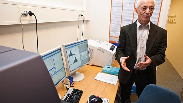
World Health Organization (WHO) expert researcher Peter Ben Embarek speaks during a press conference to wrap up a visit by an international team of experts, in the city of Wuhan, in China's Hubei Province, on February 9, 2021. (Photo by AFP)
A team of experts from the World Health Organization (WHO) investigating the origins of the COVID-19 pandemic have dismissed a controversial theory that the virus leaked from a laboratory in the Chinese city of Wuhan.
The WHO fact-finding mission, which has been in China over the past two weeks to probe the origins of the pandemic, announced during a media briefing on Tuesday that the possibility of the virus having leaked from a lab was “extremely unlikely” and did not require further study.
Peter Ben Embarek, the head of the WHO team, said Beijing had granted “full access” to all sites and personnel they had requested to conduct a comprehensive investigation.
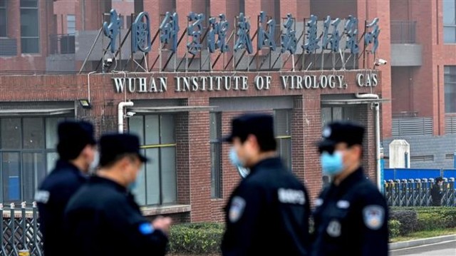
The WHO fact-finding mission arrived in Wuhan last month and visited key sites, including the Huanan seafood market, the location of the first known cluster of infections, as well as the Wuhan Institute of Virology, which was said by some to have been the actual origin of the virus.
The institute and the Chinese government had already rejected the allegation.
Since the outbreak of the epidemic, the origin of the new coronavirus has been widely discussed online, and conspiracy theories have emerged around it.
The pandemic became a political tool at the hands of former US president Donald Trump, who routinely called the pathogen “the Wuhan virus.”
WHO, intel reject Trump, Pompeo’s claim COVID-19 came from Wuhan lab
The World Health Organization says the US had provided no evidence to support US President Donald Trump’s claim that the coronavirus originated in a Chinese lab.
Trump and his associates also claimed that there had been evidence that Beijing created the new coronavirus in the medical lab in the Chinese city, even though the US intelligence agencies said they had seen no such evidence.
The coronavirus pandemic has so far claimed the lives of nearly 2,341,000 people and infected well over 107,147,260 across the world.
Wuhan lab director says virus leak claims ‘pure fabrication’
The Wuhan Institute of Virology has dismissed US claims that the coronavirus may have leaked from there.
The WHO team also said there had been no indication of the transmission of the new coronavirus in Wuhan before December 2019, when the first official cases of COVID-19 were recorded.
“In trying to understand the picture of December 2019, we embarked on a very detailed and profound search for other cases that may have been missed, cases earlier on in 2019,” said Embarek.
“And the conclusion was we did not find evidence of large outbreaks that could be related to cases of COVID-19 prior to December 2019 in Wuhan or elsewhere,” he added.
Embarek also said the team had not identified an animal source and that the virus could have been circulating in other regions before it was identified in Wuhan.
Speaking alongside Embarek, head of China’s expert panel on the outbreak, Liang Wannian, also said that early data suggested that the coronavirus could have been circulating for “several weeks” before it was identified in Wuhan.
“This indicates the possibility of the missed reported circulation in other regions,” Liang said, adding that there was “no evidence” to suggest the virus had been spreading in Wuhan before December 2019.
Press TV’s website can also be accessed at the following alternate addresses:
***
Risto Koivula
03.06.2021 00:28:22
405448
WHO: Koronaa oli Italiassa ennen kuin siitä raportoitiin Kiinassa:
" Europe
WHO Revisits Puzzling COVID-Positive Blood Samples From Pre-Pandemic Italy in Origins Hunt
19:51 GMT 02.06. 2021
by Morgan Artyukhina
The World Health Organization’s initial investigation into the origins of the COVID-19 pandemic found little reason to believe the virus leaked from a Chinese biolab, but didn’t outright dismiss the idea, either. Nonetheless, anti-China figures have continued to push the theory as being equal in validity to others, like zoonotic transfer.
WHO scientists are taking a harder look at a set of blood tests taken in Italy in October 2019 that later tested positive for antibodies against SARS-CoV-2, the virus that causes COVID-19.
On Tuesday, WHO officials confirmed they were in contact with a group of scientists in Italy who published a shocking report last November revealing that several blood tests collected as part of a lung cancer screening program months before the first cases of COVID-19 were detected in Italy had mysteriously tested positive for antibodies the body produces to fight SARS-CoV-2.
Their report, published by the Italian National Cancer Institute in its magazine Tumori Journal, found that 111 of 959 samples in the trial showed the unique spike protein SARS-CoV-2 uses to infect cells in the human body were present in the patient’s blood, and that tests of other similar coronaviruses were negative. That would mean that the patients had contracted and recovered from COVID-19 by the time of the October blood tests, signaling that COVID-19 was circulating in northern Italy in September 2019 - six months before the first cases were detected in Milan in February 2020.
"The WHO asked us if we could share the biological material and if we could re-run the tests in an independent laboratory. We accepted," Giovanni Apolone, scientific director of the Milan Cancer Institute (INT), one of the institutions responsible for the November 2020 paper, told Reuters on Monday.
According to Reuters, both positive and negative samples have been sent to Erasmus University in Rotterdam, the Netherlands, to be re-tested.
Apolone stressed that "none of the studies published so far have ever questioned the geographical origin," but likely indicate a less lethal variant of the virus has been in circulation for some time since the slew of deadlier, more virulent cases attracted medical attention in the Chinese city of Wuhan in the final days of 2019.
By February 21, 2020, the first case of COVID-19 in Europe was detected in Milan, Italy, and the city became the epicenter of Italy’s outbreak. Italy suffered the worst of the first wave of the pandemic in Europe, with a death rate as high as 7.2% - twice the death rate reported in Hubei Province, where the outbreak was first recor-ded. However, there are indications the pandemic in Italy was even worse in 2020 than reported, according to an Italian study published in January.
However, even if a milder COVID-19 infection was circulating in China earlier in 2019, it would still seem to disprove the once-discredited theory, now revived by the Biden administration, that SARS-CoV-2 escaped from the Wuhan Institute of Virology. As Sputnik has reported, the theory was spread by former US President Donald Trump and right-wing anti-China figures close to him in early 2020 as the pandemic spiraled out of control in the US and his administration began looking for someone else to blame for it.
Last week, WHO spokesperson Fadela Chaib told a United Nations briefing the agency was preparing a proposal on a second phase of its investigation into the origins of SARS-CoV-2, but she provided no specific timeline. The first phase, con-ducted in January and February and released in March,yielded no concrete results, but found a zoonotic exchange - that humans caught it from an infected animal - more likely than that the virus escaped from the Wuhan lab. They also suggested an international military sports tournament the city hosted in October 2019 be investigated as an additional possible vector.
Some scientists and political figures intent on pushing the “lab leak” theory have claimed Beijing denied the WHO scientists adequate access to data from the Wu-han virology lab, with more than a dozen US allies issuing a statement demanding openness, but scientists on the WHO team have pushed back, saying their efforts weren’t impeded by Chinese officials, and that the level of openness being deman-ded of China is one no Western country would accept if it were made of them.
Also last week,US President Joe Biden ordered an intelligence report on the origins of COVID-19, "including whether it emerged from human contact with an infected animal or from a laboratory accident,” to be delivered in 90 days. In response, the Global Times called for the US biolab at Fort Detrick, Maryland, to be investigated as well. "
Risto Koivula
03.06.2021 23:45:58
405453
WHO: Koronaa oli Italiassa ennen kuin siitä raportoitiin Kiinassa:
Lainaus: riiviö, 03.06.2021 17:41:37, 405450
Linkki riittää mainiosti ja sitten se oma asia, jos sitä on ja jos sen osaa kirjottaa.
Tulipa sanotuksi. Helpotti. En ole täällä ollenkaan moderointitehtävissä.
RK: Linkit menee kiinni ja tunnusten taakse. Tätä asiaa on syytä voida seurata 10:nkin vuoden päästä.
Risto Koivula
04.06.2021 02:36:34
405456
HEWOWWITTUA, YLE!!! SITÄ *EI* OLE "MUOKATTU"!!!
https://yle.fi/uutiset/3-11963398
Sähköpostit paljastavat: Tutkija varoitti varhain Yhdysvaltain korona-asiantuntija Anthony Faucia, että virusta on mahdollisesti muokattu
Buzzfeedin julkaisemien sähköpostien perusteella Faucilla oli syytä pitää laboratoriovuotoa uskottavana vaihtoehtona koronaviruksen alkuperälle.
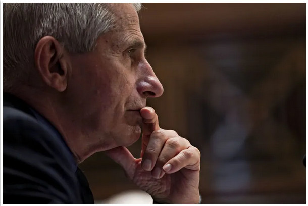
Anthony Fauci
Kansallisen allergia- ja infektiotautien instituutin pääjohtaja Anthony Fauci toimii Valkoisen talon korona-asiantuntijana.Stefani Reynolds / EPA
3.6. 16:46
Yhdysvaltalaisen tartuntatautien tutkimuslaitoksen johtaja lähetti Valkoisen talon ko-rona-asiantuntija Anthony Faucille tammikuun 2020 lopussa sähköpostin, jossa hän kertoi, että pieni osa koronaviruksen perimästä näyttää mahdollisesti muokatulta.
Asia selviää Faucin sähköposteista, jotka Buzzfeed-sivusto sai haltuunsa tietopyyn-nön kautta. Myös Washington Post (siirryt toiseen palveluun) on hankkinut Faucin sähköposteja itselleen.
Buzzfeed on ladannut yli 3 200 sivua sähköposteja nettiin kaikkien tarkasteltaviksi. (siirryt toiseen palveluun) Viestit ovat tammi-kesäkuulta 2020.
Fauci tyrmäsi julkisesti keväällä 2020 teorian siitä, että pandemian aiheuttanut Sars-Cov-2 -koronavirus olisi peräisin kiinalaisesta laboratoriosta.
Buzzfeedin saamien sähköpostien perusteella hänellä kuitenkin näytti olleen syytä pitää laboratoriovuotoa uskottavana vaihtoehtona viruksen alkuperälle.
Tutkijat kiinnittivät huomiota viruksen erityispiirteisiin
Immunologian ja mikrobiologian professori Kristian G. Andersen kirjoitti Faucille 31. tammikuuta 2020 lähettämässään sähköpostissa, että hän ja kolme kollegaa katsovat viruksen geeniperimän vaikuttavan epäjohdonmukaiselta evoluutioteoriaan liittyvien odotusten kannalta.
Hän toimii Scrippsin tutkimusinstituutin Andersen Labin johtajana.
– Viruksen epätavalliset erityispiirteet muodostavat todella pienen osan geeniperi-mästä (<0,1%), joten kaikkia sekvenssejä täytyy tarkastella hyvin tarkasti, jotta voi nähdä, että osa ominaisuuksista (mahdollisesti) näyttää muokatuilta, Andersen kirjoitti.
Andersen kertoi, että evoluutiopuussa virus näyttää täysin normaalilta ja se näyttäisi olevan peräisin lepakoista.
Andersen lisäsi, että asia vaati lisää analyyseja ja heidän mielipiteensä voivat vielä muuttua.
Lyhyessä vastausviestissään Fauci kiittää Andersenia ja sanoo, että keskustellaan pian puhelimitse. Hän ei tuonut tutkijoiden näkemystä julki.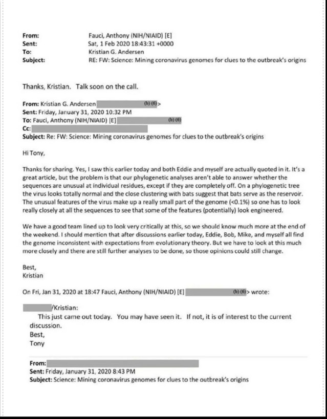
Andersen pitää nyt laboratoriovuotoa epätodennäköisenä
Myöhemmin professori Kristian G. Andersenin mielipide muuttui.
Hänen ryhmänsä julkaisi maaliskuussa 2020 Nature-lehdessä (siirryt toiseen palve-luun) tutkimuksen, jonka mukaan on epätodennäköistä, että Sars-Cov-2 on syntynyt Sarsin kaltaisen koronaviruksen muokkauksen tuloksena.
Huhtikuussa 2021 Andersen avasi Twitterissä näkemystään, miksi hän pitää labora-toriovuotoa epätodennäköisenä. Andersen kertoi, että he etsivät merkkejä viruksen muokkauksesta laboratoriossa, mutta eivät löytäneet siihen viittaavia todisteita.
Andersen sanoi, että jos laboratoriovuototeoria pitäisi paikkansa, se vaatisi suurta peittelyä ja valehtelua monilta arvostetuilta tutkijoilta Kiinassa ja muualla.
– Todistaako raportti varmasti, että laboratoriovuotoa ei tapahtunut? Ei todista. Tarkoittaako se, että meidän ei tulisi harkita laboratoriovuodon mahdollisuutta? Ei tarkoita. Sen sijaan rapotin johtopäätös on, että se on erittäin epätodennäköinen, koska muut hypoteesit ovat paljon todennäköisempiä, Andersen kirjoitti Twitterissä.
Lisäksi Andersen sanoi, että pandemian alkuperä vaatii lisää avoimia tutkimuksia. Hän arvioi, että ne todennäköisesti kestävät vuosia.
"Salaliittoteoria" kiinnosti Faucin pomoa
Yhdysvaltain terveysviraston NIH:n johtaja Francis Collins lähetti 16. huhtikuuta 2020 Faucille ja muutamalle muulle terveysviranomaiselle sähköpostin otsikolla "salaliitto kerää vauhtia".
Fauci toimii terveysviraston alaisen kansallisen allergia- ja infektiotautien instituutin pääjohtajana. Collins on Faucin pomo.
Collinsin viestissä oli linkki Mediaite-sivuston uutiseen, joka käsitteli Fox News-kanavalla käytyä keskustelua viruksen mahdollisesta karkaamisesta wuhanilaisesta laboratoriosta.
Collinsin viestin sisältö on mustattu Buzzfeedin saamista sähköposteista yllä mainittua linkkiä lukuunottamatta. Fauci vastasi Collinsille, mutta myös hänen vastauksensa on peitetty.
Wuhanilaislaboratorion kumppanilta kiitokset
Pari päivää myöhemmin, 18. huhtikuuta 2020, Fauci sai sähköpostin brittiläiseltä eläintieteilijältä Peter Daszakilta, joka johtaa EcoHealth Alliance -tutkimusjärjestöä.
Daszak kirjoitti haluavansa kiittää Faucia henkilökuntansa ja yhteistyötahojensa puo-lesta siitä, että Fauci oli julkisesti sanonut tieteellisen todistusaineiston tukevan sitä, että Covid-19 on tarttunut luonnollisella tavalla lepakoista ihmisiin eikä ole karannut Wuhanin virologisen instituutin laboratoriosta.
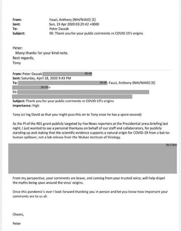
Suuri osa viestistä on peitetty. Lopuksi Daszak kehuu, että Faucin rohkeat kommentit auttavat kumoamaan väitteitä viruksen alkuperästä.
Fauci vastasi, että paljon kiitoksia ystävällisestä viestistäsi.
Daszakin johtama EcoHealth Alliance on tehnyt vuosia läheistä yhteistyötä Wuhanin virologisen instituutin kanssa koronavirusten tutkimuksessa.
Monet ihmiset kyselevät nyt Twitterissä Daszakilta, mitä viestin peitetyssä osassa sanottiin. Toiset syyttävät häntä murhaajaksi, koska he katsovat hänen olevan osasyyllinen pandemiaan.
Laboratorioalkuperä noussut vaihtoehdoksi viime aikoina (Valetta, pelkkää hörölöröä)
Fauci sanoi viime kuussa lainsäätäjille,että Yhdysvaltain terveysvirasto oli myöntänyt 600 000 dollarin apurahan Wuhanin virologiselle instituutille. Rahoitus oli ohjattu kiinalaislaboratoriolle juuri EcoHealth Alliancen kautta.
Viisivuotisen apurahan avulla oli tarkoitus tutkia koronavirusten tarttumista lepakois-ta ihmisiin, jotta tutkijat ymmärtäisivät paremmin 2000-luvun alkupuolen Sars-epidemiaa. Trumpin hallinto keskeytti rahoituksen viime vuonna.
Peter Daszak kertoi joulukuussa 2019 podcast-haastattelussa (siirryt toiseen palveluun), että koronaviruksia voi muokata melko helposti laboratoriossa.
Daszak kuului Maailman terveysjärjestö WHO:n tutkimusryhmään, joka yritti selvittää koronapandemian lähdettä Wuhanissa. Hän on myös jäsen tieteellisen Lancet-julkaisun ryhmässä, joka selvittää pandemian alkuperää.
Daszakia on arvosteltu eturistiriidasta kaksoisroolinsa vuoksi. Hän on kutsunut viruksen mahdollista karkaamista wuhanilaislaboratoriosta salaliittoteoriaksi.
Viruksen mahdollinen laboratorioalkuperä on noussut varteen otettavaksi vaihtoehdoksi viime aikoina.
Wall Street Journal -lehti (siirryt toiseen palveluun) uutisoi toukokuussa, että kolme Wuhanin virologisen instituutin työntekijää joutui marraskuussa 2019 sairaalaan, koska heillä oli koronatartuntaan viittaavia oireita.
Fauci kannattaa lisätutkimuksia
Anthony Fauci sanoi toukokuussa senaatille, että hän ja suurin osa tieteellisestä yhteisöstä pitää todennäköisimpänä skenaariona, että Sars-Cov-2 on peräisin luonnosta, mutta kukaan ei voi olla siitä täysin varma.
CNN:n mukaan (siirryt toiseen palveluun) Fauci sanoi haastattelussa, että hän ei ole vakuuttunut siitä, että virus olisi syntynyt luonnollisesti. Fauci sanoi kannattavansa tutkimusta siitä, mitä Kiinassa tapahtui.
Presidentti Joe Biden on määrännyt Yhdysvaltain tiedustelupalvelut selvittämään, pääsikö virus karkuun kiinalaisesta laboratoriosta.
Asian selvittämistä hankaloittaa se, että Kiina ei ole suostunut avoimeen kansainväli-seen tutkintaan. Kiina on kiistänyt viruksen karkaamisen laboratoriosta ja on kutsunut väitettä salaliittoteoriaksi.
Lue lisää:
Yhdysvallat rahoitti virustutkimusta wuhanilaisessa laboratoriossa – osa republikaaneista vaatii nyt korona-asiantuntija Anthony Faucin eroa
Jani Kaaron kolumni: Entäpä jos koronavirus onkin laboratoriokarkulainen Wuhanin viruslaboratoriosta? (täyttä paskaa)
***
ForbesLifestyle Travel Editors' Pick
" Controversial Coronavirus Lab Origin Claims Dismissed By Experts

Wuhan Institute of Virology is one of China's leading research centres and has been the subject of ... [+]
AFP via Getty ImagesJune 8: This article has been substantially updated to reflect criticism of the published study, along with the general scientific consensus on Covid-19. It also clarifies Sørensen’s financial interest in the development of the Biovacc-19 coronavirus vaccine. This context did not appear in the original post. June 10: Updated with additional comment from Gunnveig Grødeland.
Sir Richard Dearlove, who was head of MI6 from 1999 to 2004, told the Daily Telegraph that Sørensen and Dalgleish’s research shows that the pandemic may have started at the Wuhan Institute of Virology. He added that he thought it unlikely to have been released deliberately, but that China had clearly tried to cover up the release.
However, intelligence sources from Britain’s MI5 dismissed the idea as “rumor and conspiracy,” according to the Times of London. Dearlove was previously criticized in the Chilcot Inquiry into the Iraq War for promoting “flawed intelligence.”
Since the coronavirus took hold in the United States, senior officials in the Trump administration have amplified rumors that the virus emerged from a virology lab in Wuhan. However, public health researchers have traced the earliest recorded cases of the virus to an animal market in the city.
Scientific claims questioned
Sørensen and Dalgleish’s work contradicts the international scientific consensus that although the coronavirus pandemic originated in Wuhan, there is no evidence that it had been artificially engineered.
An analysis of the first 41 Covid-19 patients published in medical journal the Lancet found that in 27 cases, there had been direct exposure to the Wuhan market, although not with the first known case. The World Health Organization has since released guidance to those working in and visiting such markets, to reduce animal-human transmission of emerging pathogens.
The report’s authors also claim the lack of mutation in the virus since its discovery, suggests it was already fully adapted to humans. However, there have been several published studies noting evolution and mutation among SARS-CoV-2 strains.
Sørensen told Norwegian broadcaster NRK that the virus has properties that differ greatly from SARS, another coronavirus, and have never been detected in nature. He claimed that China and the United States have collaborated for many years on coronavirus research through "gain of function" studies, in which the pathogenicity or transmissibility of potential pandemic pathogens can be enhanced in order to understand them better.
Examples found in other viruses
Gunnveig Grødeland, vaccine researcher at the University of Oslo, is one of the scientists voicing their disagreement with Sørensen. She explains that what Sørensen referred to as "inserted sequences" can enable the development of a more serious disease, but this is not unusual in nature: "Examples can be found in other viruses including subtypes of influenza (including "bird flu"), HIV, and several human coronaviruses (MERS, OC43, HKU1)." Grødeland also says that Sørensen's paper offered no biological confirmation on the relevance of positively charged patches.
Other leading voices in the scientific community and fight against the pandemic have dismissed claims that the coronavirus was man-made. Anthony Fauci, director of U.S. National Institute of Allergy and Infectious Diseases, has stated there is no scientific evidence for the claims, while leading researchers from the World Health Organization and Galveston National Laboratory also dismissed the rumours.
The report was allegedly previously rejected
The Telegraph claimed that the study was initially rejected by several academic journals, including "Nature" and "Journal of Virology," which indicates they considered the article unsuitable for publication. Sørensen claimed the original study tied the vaccine development to the origin research, and that they have since split the research. He claimed a second study with more details on the origin of the virus will be published soon.
The report published in the Quarterly Review of Biophysics explains the rationale for the development of Biovacc-19, a candidate vaccine for COVID-19 that is now in advanced preclinical development. Sørensen has a financial interest in Immunor, the Norwegian company behind the vaccine.
No, COVID-19 Coronavirus Was Not Bioengineered. Here’s The Research That Debunks That Idea (Forbes)
Fauci, Top U.S. General Throw Cold Water On Trump’s Coronavirus Claim (Forbes)
The Controversial Rumor COVID-19 Originated In A Wuhan Lab Creeps Into The GOP Mainstream (Forbes)
A Timeline Of The COVID-19 Wuhan Lab Origin Theory (Forbes)

***
Haistapaskantiede-Birger Sørensenin "todistus":
https://www.nrk.no/norge/norsk-forsker-skaper-strid-om-virusets-opphav_-_-dette-viruset-har-ikke-en-naturlig-opprinnelse-1.15043634
Norsk forsker skaper strid om virusets opphav: – Dette viruset har ikke en naturlig opprinnelse
Sekvenser i koronavirusets overflate viser at det ikke kommer fra naturen, men trolig er utviklet av kinesiske og amerikanske forskere. Det mener den norske vaksineforskeren Birger Sørensen. Han får støtte av britenes tidligere spionsjef.
IKKE NATURLIG: Vaksineforsker Birger Sørensen mener viruset SARS-CoV-2 ikke kommer fra naturen. Bildet er fra 2009. Foto: Støtvig Alf Øystein
Publisert 7. juni 2020 kl. 19:49 Oppdatert 8. juni 2020 kl. 18:34
Artikkelen er mer enn to år gammel.
Rettelse: I en tidligere versjon av saken stod det at studien påviser at sekvenser i koronavirusets spike-protein ser ut til å være kunstig satt inn. Denne konklusjonen er bestridt av norske forskere, og omtales heller ikke i forskningsartikkelen. Denne påstanden var basert på uttalelser fra Sørensen til NRK. Saken burde hatt flere relevante kilder som vurderte Sørensens uttalelser og forskning. Dette har NRK nå publisert i en egen sak.
https://usrtk.org/scientific-papers-on-the-origins-of-sars-cov-2/
" Scientific papers on the origins of SARS-CoV-2
Here is a list of papers published in peer-reviewed scientific journals on the origins of SARS-CoV-2, the virus that causes COVID-19.
Science. The Huanan Seafood Wholesale Market in Wuhan was the early epicenter of the COVID-19 pandemic. By Michael Worobey et al. July 26, 2022.
Science. The molecular epidemiology of multiple zoonotic origins of SARS-CoV-2. By Jonathan E. Pekar et al. July 26, 2022.
Nature. Wuhan market was epicentre of pandemic’s start, studies suggest. By Amy Maxmen. February 27, 2022.
Zenodo. The Huanan market was the epicenter of SARS-CoV-2 emergence. Michael Worobey et al. February 26, 2022.
Zenodo. SARS-CoV-2 emergence very likely resulted from at least two zoonotic events. Jonathan Pekar et al. February 26, 2022.
Nature Portfolio. Surveillance of SARS-CoV-2 in the environment and animal samples of the Huanan Seafood Market. George Gao et al. February 25, 2022.
Nature Portfolio. Coronaviruses with a SARS-CoV-2-like receptor-binding domain allowing ACE2-mediated entry into human cells isolated from bats of Indochinese peninsula. Sarah Temmam et al. September 17, 2021. Under review.
Nature.Origins of SARS-CoV-2: window is closing for key scientific studies. August 25, 2021.
Cell. The Origins of SARS-CoV-2: A Critical Review (pre-proof). Edward C. Holmes et al. August 20, 2021.
Science. The animal origin of SARS-CoV-2. Spyros Lytras,Wei Xia,Joseph Hughes,Xiaowei Jiang andDavid L. Robertson. August 17, 2021.
mBio. Can Science Help Resolve the Controversy on the Origins of the SARS-CoV-2 Pandemic? Arturo Arturo Casadevall, Susan R. Weiss and Michael Imperiale. August 2, 2021.
Independent Science News. Phylogeographic Mapping of Newly Discovered Coronaviruses Pinpoints the Direct Progenitor of SARS-CoV-2 as Originating from Mojiang, China. Jonathan Latham and Allison Wilson. August 2, 2021.
Frontiers in Public Health. Lethal Pneumonia Cases in Mojiang Miners (2012) and the Mineshaft Could Provide Important Clues to the Origin of SARS-CoV-2. Alex C. Speciale. July 13, 2021.
Medium. A response to “The Origins of SARS-CoV-2: A Critical Review.” Alina Chan. July 12, 2021.
Nature Scientific Reports. In silico comparison of SARS-CoV-2 spike protein-ACE2 binding affinities across species and implications for virus origin. Sakshi Piplani, Puneet Kumar Singh, David A. Winkler, Nikolai Petrovsky. June 24, 2021. doi: https://doi.org/10.1038/s41598-021-92388-5
bioRxiv. Recovery of deleted deep sequencing data sheds more light on the early Wuhan SARS-CoV-2 epidemic. Jesse Bloom. June 22, 2021. doi: https://doi.org/10.1101/2021.06.18.449051
In Vivo. On the Origin of SARS-CoV-2: Did Cell Culture Experiments Lead to Increased Virulence of the Progenitor Virus for Humans? Bernd Kaina. May 2021, 35 (3) 1313-1326; DOI: https://doi.org/10.21873/invivo.12384
Infectious Diseases & Immunity. Origins of SARS-CoV-2: Focusing on Science. Zhengli Shi. April 2021 – Volume 1, Issue 1, p.3-4 doi: 10.1097/ID9.0000000000000008
Environmental Chemistry Letters. Should we discount the laboratory origin of COVID-19? Rossana Segreto, Yuri Deigin, Kevin McCairn, Alejandro Sousa, Dan Sirotkin, Karl Sirotkin, Jonathan J. Couey, Adrian Jones & Daoyu Zhang. March 25, 2021.
Environmental Chemistry Letters. Tracing the origins of SARS-COV-2 in coronavirus phylogenies: a review. Erwan Sallard, José Halloy, Didier Casane, Etienne Decroly and Jacques van Helden. February 4, 2021. doi: https://doi.org/10.1007/s10311-020-01151-1
Zenodo. A Bayesian analysis concludes beyond a reasonable doubt that SARS-CoV-2 is not a natural zoonosis but instead is laboratory derived. Dr. Steven Quay. January 29, 2021.
Nature. Addendum: A pneumonia outbreak associated with a new coronavirus of probable bat origin. Peng Zhou, Xing-Lou Yang, Xian-Guang Wang, Ben Hu,…and Zheng-Li Shi. November 17, 2020. https://doi.org/10.1038/s41586-020-2951-z
BioEssays. The genetic structure of SARS‐CoV‐2 does not rule out a laboratory origin. Rossana Segreto and Yuri Deigin. November 17, 2020. https://doi.org/10.1002/bies.202000240
Zenodo. Where Did the 2019 Coronavirus Pandemic Begin and How Did it Spread? The People’s Liberation Army Hospital in Wuhan China and Line 2 of the Wuhan Metro System Are Compelling Answers. Steven Carl Quay. October 28, 2020. doi: 10.5281/zenodo.4119262
Frontiers in Public Health. Lethal pneumonia cases in Mojiang miners (2012) and the mineshaft could provide important clues to the origin of SARS-CoV-2. Monali Rahalkar and Rahul Bahulikar. September 17, 2020. doi: 10.3389/fpubh.2020.581569
Journal of Medical Virology. Questions concerning the proximal origin of SARS-CoV-2. Murat Seyran, Damiano Pizzol, Parise Adadi…and Adam M. Brufsky. September 3, 2020. doi: https://doi.org/10.1002/jmv.26478
BioEssays. Might SARS‐CoV‐2 have arisen via serial passage through an animal host or cell culture? Karl Sirotkin and Dan Sirotkin. August 12, 2020. https://doi.org/10.1002/bies.202000091
bioRxiv. SARS-CoV-2 is well adapted for humans. What does this mean for re-emergence? Shing Hei Zhan, Benjamin E. Deverman, Yujia Alina Chan. May 2, 2020. doi: https://doi.org/10.1101/2020.05.01.073262
Nature Medicine. The proximal origin of SARS-CoV-2. Kristian G. Andersen, Andrew Rambaut, W. Ian Lipkin, Edward C. Holmes, Robert F. Garry. April 2020. Volume 26, pages 450-455.
Antiviral Research. The spike glycoprotein of the new coronavirus 2019-nCoV contains a furin-like cleavage site absent in CoV of the same clade. Bruno Coutard et al. February 10, 2020. DOI: 10.1016/j.antiviral.2020.104742
Nature. A pneumonia outbreak associated with a new coronavirus of probable bat origin. Peng Zhou, Xing-Lou Yang, Xian-Guang Wang, Ben Hu,…and Zheng-Li Shi. February 3, 2020. 579(7798): 270-273. doi:10.1038/s41586-020-2012-7
Preprint. The possible origins of 2019-nCoV coronavirus. Botao Xiao. February 2020. doi: 10.13140/RG.2.2.21799.29601
The Lancet. Clinical features of patients infected with 2019 novel coronavirus in Wuhan, China. Chaolin Huang et al. January 30, 2020. Volume 395: 497–506.
Scientific Reports. Animal sales from Wuhan wet markets immediately prior to the COVID-19 pandemic. Xiao Xiao et al. June 7, 2021. Sci Rep 11, 11898 (2021). https://doi.org/10.1038/s41598-021-91470-2
Minerva. The evidence which suggests that this is no naturally evolved virus: A reconstructed historical aetiology of the SARS-CoV-2 spike. Birger Sørensen, Angus Dalgleish & Andres Susrud. July 1, 2020.
ResearchGate. Is considering a genetic-manipulation origin for SARS-CoV-2 a conspiracy theory that must be censored? Rossana Segreto and Yuri Deigin. April 2020. DOI: 10.13140/RG.2.2.31358.13129/1
Preprints. Major concerns on the identification of bat coronavirus strain RaTG13 and quality of related Nature paper. Xiaoxu Lin, Shizhong Chen. June 5, 2020. 2020060044. doi: 10.20944/preprints202006.0044.v1
Preprints. The abnormal nature of the fecal swab sample used for NGS analysis of RaTG13 genome sequence imposes a question on the correctness of the RaTG13 sequence. Monali Rahalkar and Rahul Bahulikar. August 11, 2020. doi: 10.20944/preprints202008.0205.v1
OSF Preprints. COVID-19, SARS and bats coronaviruses genomes unexpected exogeneous RNA sequences. Jean-Claude Perez and Luc Montagnier. April 25, 2020. doi:10.31219/osf.io/d9e5g
Zenodo. HIV man-manipulated coronavirus genome evolution trends. Jean-Claude Perez and Luc Montagnier. August 2, 2020.
Emerging Microbes & Infections. HIV-1 did not contribute to the 2019-nCoV genome. Xiao Chuan, Li Xiaojun, Liu Shuying, Sang Yongming, Gao Shou-Jiang and Gao Feng. 2020. 9(1): 378-381. doi: 10.1080/22221751.2020.1727299
Nature. Identifying SARS-CoV-2-related coronaviruses in Malayan pangolins. Tommy Tsan-Yuk Lam, Na Jia, Ya-Wei Zhang, Marcus Ho-Hin Shum, Jia-Fu Jiang, Hua-Chen Zhu, Yi-Gang Tong, Yong-Xia Shi, Xue-Bing Ni, Yun-Shi Liao, Wen-Juan Li, Bao-Gui Jiang, Wei Wei, Ting-Ting Yuan, Kui Zheng, Xiao-Ming Cui, Jie Li, Guang-Qian Pei, Xin Qiang, William Yiu-Man Cheung, Lian-Feng Li, Fang-Fang Sun, Si Qin, Ji-Cheng Huang, Gabriel M. Leung, Edward C. Holmes, Yan-Ling Hu, Yi Guan & Wu-Chun Cao. March 26, 2020. doi: https://doi.org/10.1038/s41586-020-2169-0
PLoS Pathogens. Are pangolins the intermediate host of the 2019 novel coronavirus (SARS-CoV-2)? Ping Liu, Jing-Zhe Jiang, Xiu-Feng Wan, Yan Hua, Linmiao Li, Jiabin Zhou, Xiaohu Wang, Fanghui Hou, Jing Chen, Jiejian Zou, Jinping Chen. May 14, 2020. doi: https://doi.org/10.1371/journal.ppat.1008421
Nature. Isolation of SARS-CoV-2-related coronavirus from Malayan pangolins. Kangpeng Xiao, Junqiong Zhai, Yaoyu Feng, Niu Zhou, Xu Zhang, Jie-Jian Zou, Na Li, Yaqiong Guo, Xiaobing Li, Xuejuan Shen, Zhipeng Zhang, Fanfan Shu, Wanyi Huang, Yu Li, Ziding Zhang, Rui-Ai Chen, Ya-Jiang Wu, Shi-Ming Peng, Mian Huang, Wei-Jun Xie, Qin-Hui Cai, Fang-Hui Hou, Wu Chen, Lihua Xiao & Yongyi She. May 7, 2020. doi: https://doi.org/10.1038/s41586-020-2313-x
Current Biology. Probable Pangolin Origin of SARS-CoV-2 Associated with the COVID-19 Outbreak. Tao Zhang, Qunfu Wu, Zhigang Zhang. March 19, 2020. doi: https://doi.org/10.1016/j.cub.2020.03.022
bioRxiv. Single source of pangolin CoVs with a near identical Spike RBD to SARS-CoV-2. Yujia Alina Chan and Shing Hei Zhan. October 23, 2020. doi: https://doi.org/10.1101/2020.07.07.184374
Infection, Genetics and Evolution. COVID-19: Time to exonerate the pangolin from the transmission of SARS-CoV-2 to humans. Roger Frutos, Jordi Serra-Cobo, Tianmu Chen and Christian A. Devaux. Volume 84, October 2020, 104493. https://doi.org/10.1016/j.meegid.2020.104493
bioRxiv. No evidence of coronaviruses or other potentially zoonotic viruses in Sunda pangolins (Manis javanica) entering the wildlife trade via Malaysia. Jimmy Lee, Tom Hughes, Mei-Ho Lee, Hume Field, Jeffrine Japning Rovie-Ryan, Frankie Thomas Sitam, Symphorosa Sipangkui, Senthilvel K.S.S. Nathan, Diana Ramirez, Subbiah Vijay Kumar, Helen Lasimbang, Jonathan H. Epstein, Peter Daszak. June 19, 2020. doi: https://doi.org/10.1101/2020.06.19.158717
Cell. A genomic perspective on the origin and emergence of SARS-CoV-2. Yong-Zhen Zhang, Edward C. Holmes. April 2020 181(2):223-227. doi: 10.1016/j.cell.2020.03.035.
Current Biology. A novel bat coronavirus closely related to SARS-CoV-2 contains natural insertions at the S1/S2 cleavage site of the spike protein. Hong Zhou, Xing Chen, Tao Hu, Juan Li, Hao Song, Yanran Liu, Peihan Wang, Di Liu, Jing Yang, Edward C. Holmes, Alice C. Hughes, Yuhai Bi, and Weifeng Shi. June 8, 2020. 30: 2196-2203. doi: https://doi.org/10.1016/j.cub.2020.05.023
aRxiv. The bat coronavirus RmYN02 is characterized by a 6-nucleotide deletion at the S1/S2 junction, and its claimed PAA insertion is highly doubtful. Yuri Deigin and Rossana Segreto. December 1, 2020.
Zenodo. Unusual features of the SARS-CoV-2 genome suggesting sophisticated laboratory modification rather than natural evolution and delineation of its probable synthetic route. Li-Meng Yan, Shu Kang, Jie Guan, and Shanchang Hu. September 14, 2020. doi: 10.5281/zenodo.4028829
Johns Hopkins Center for Health Security. In Response: Yan et al Preprint Examinations of the Origin of SARS-CoV-2. Kelsey Lane Warmbrod, Rachel M. West, Nancy D. Connell and Gigi Kwik Gronvall. September 21, 2020.
Zenodo. Proposed SARS-CoV-2 Spillover During 2019 Review of Samples from a Mineshaft in Mojiang, Yunnan Province, China. Anonymous. September 14, 2020. doi: 10.5281/zenodo.4029544
ResearchGate. An investigation into the WIV databases that were taken offline. Billy Bostickson et al. February 2021.
ResearchGate. Wuhan Institute of Biological Products Co. Rodolphe de Maistre, Gilles Demaneuf and Billy Bostickson. March 2021.
Zenodo. 1. Proposed Forensic Investigation of Wuhan Laboratories. Billy Bostickson and Yvette Ghannam. March 2021. DOI: 10.5281/zenodo.4640383
ResearchGate. 2. Investigation of RaTG13 and the 7896 Clade. Billy Bostickson and Yvette Ghannam. March 2021. DOI: 10.13140/RG.2.2.22382.33607
ResearchGate. 3. Wuhan Laboratories, Bat Research and Biosafety. Billy Bostickson and Yvette Ghannam. April 2021. DOI: 10.13140/RG.2.2.32006.29761
***
Hesari-tyhmentymän Aamukakka-lehtikin lopulta ottaa lusikan kauniiseen käteen, että todisteet koroviruksen luonnollisesta alkuperästä ovat ehdottomat, eikä Dalgleishin ja Sörensenin eikä muitakaan "keinotekoituusteorioita" enää selvitellä. Lainaus jutusta:
" *** Toki on myös muita teorioita. Esimerkiksi se, että karkuun päässyt virus olisi itse valmistettu kiinalaislaboratoriossa. Kiinassa taas on esitetty, että virus olisi valmistettu eräässä Yhdysvalloissa sijaitsevassa biolaboratoriossa. Nämä salaliittoteoriat voidaan jättää huomioimatta. *** "
Nyt vaaditaan tutkittavaksi viruksen etenemisREITTIÄ luonnosta ihmiskunnan pandemiaksi. Siinä asiassa pelataan tiedolla, jota ei voida ehdottomasti todistaa todeksi tai epätodeksi kuten itse viruksen alkuperä. Jotkut trooppiset lepakot vaivat lentää vaikka maapallom ympäri monsuunituulten mukana ja tartuttaa viruksiaan myös aivan muihin nisäkäsryhmiin.
https://www.aamulehti.fi/ulkomaat/art-2000009452421.html
" Vieläkin kiistellään, mistä miljoonia tappanut koronapandemia sai alkunsa – Tällä hetkellä vastakkain ovat nämä kaksi teoriaa
Koronaviruksen aiheuttama covid-19-tauti julistettiin pandemiaksi kolme vuotta sitten, mutta vieläkin kiistellään siitä, mistä miljoonia ihmisiä surmannut virus sai alkunsa.

Japanissa maanantaista alkaen maskia ei ole tarvinnut enää käyttää esimerkiksi julkisissa liikennevälineissä muutoin kuin ruuhka-aikoina. Moni jatkoi maskin käyttöä siitä huolimatta myös kadulla. Kuva: Kim Kyung Koon / Reuters
-
Risto Koivula tuota ekaa kommenttiasi en ymmärtänyt. Mutta olet sitä mieltä, että on OK kehitellä viruksista vaarallisempia ihmiselle? Eli otetaan jokin virus luonnosta joka leviää ilmateitse ja lisätään sille kyky tunkeutua ihmissoluihin jota sillä luonnollisesti ei ole?
Risto Koivula: MITÄÄN SELLAISTA PIIKKIPROTEIINIA MIHINKÄÄN LEPAKKOJEN KORONAVIRUKSEEN EI OLE LISÄTTY.
https://www.pnas.org/doi/10.1073/pnas.2211107119
" SARS-CoV-2 furin cleavage site was not engineered

Numerous features of SARS-CoV-2 FCS demonstrate that it was not engineered to mimic human ENaC:
References
" Financial analyst David Martin misrepresents studies and patent applications to promote the baseless claim that SARS-CoV-2 was developed as a bioweapon


Factually inaccurate: Old patent applications for coronavirus vaccines and vectors cited by Martin are for animal coronaviruses. None of them is related to SARS-CoV-2 nor do they prove that the virus was developed as a bioweapon.
FULL CLAIM: “SARS is the research developed by humans, weaponizing a life system model to actually attack human beings, and they patented it in 2002.”; “The intent was to get the world to accept a universal vaccine template, and the intent was to use coronavirus to get there”
REVIEW
In May 2023, a video featuring financial analyst David Martin went viral on social media platforms, gathering millions of views on Facebook, Instagram, Twitter, and TikTok (examples here, here, here, and here). In it, Martin claimed that SARS-CoV-2 was an act of “premeditated domestic terrorism” and that the virus is a weapon that was engineered through decades of research with the objective of “get[ting] the world to accept a universal vaccine template”.
Martin’s 21-minute speech was only the beginning of a nine-hour event called “Inter-national COVID-19 Summit III” that was held in a room of the European Parliament in Brussels on 3 May 2023. As featured speakers, the event gathered a group of well-known COVID-19 misinformation actors, including Robert Malone, Ryan Cole, Byram Bridle, Christian Perronne, and Pierre Kory, president of the fringe medical group Front Line COVID-19 Critical Care Alliance (FLCCC).
A three-part video recording of the event was uploaded on Rumble, nd video excerpts were widely spread online, aided by anti-vaccine, COVID-19 skeptic, and conspiracy groups and individuals who shared them. Among all the speakers, Martin’s speech attracted much attention online, receiving shares from FLCCC, the conspiracy website Infowars, and the anti-vaccine group Children’s Health Defense, as well as many social media users.
However, Martin’s claims are all unsubstantiated, contain numerous inaccuracies, and are based on a grossly distorted interpretation of historical coronavirus research that we will analyze in detail below.
The place where the summit took place and the summit’s use of the European Parliament’s logo was also misleading, lending a veneer of legitimacy to claims that the speakers had testified to the European Parliament. However, this wasn’t the case. Firstly, the event doesn’t appear in the list of official events on the European Parliament website.
Secondly, the event wasn’t held at the Hemicycle, where the 705 Members of the European Parliament (MEPs) gather during plenary sessions. Instead, it took place in one of the rooms available to MEPs, which was possible because the Summit was hosted by five MEPs, Cristian Terhes, Christine Anderson, Ivan Vilibor Sinčić, Francesca Donato, and Mislav Kolakušić, all of them known COVID-19 skeptics.
Thirdly, the European assembly clearly explained to CheckNews—a fact-checking section of the French newspaper Libération—that the European Parliament “didn’t organize or finance” this event, although some MEPs took part, “exercising their freedom of mandate”.
Claim 1 (Inaccurate and Misleading):
“coronavirus as a model of a pathogen was isolated in 1965. Coronavirus was identified in 1965 as one of the first infectious replicatable [sic] viral models that could be used to modify a series of other experiences of human condition.”
While it is unclear what Martin meant with “a model of pathogen”, the first corona-virus wasn’t isolated in 1965 but in 1936. That year, Beach and Schalm isolated the pathogen that caused avian bronchitis in newborn chickens, which symptoms had been described five years earlier, and determined that the disease was caused by a virus [1].
It still took 30 years to identify the first human coronavirus, 229E, discovered by Do-rothy Hamre at the University of Chicago in 1962 when she analyzed tissue cultures of students with colds[2]. Around the same time, two research teams, one led by Da-vid Tyrrell in England and another by Ken McInstosh in the U.S., independently iso-lated two additional human coronaviruses, called B814 and OC43, respectively [3,4].
However, Martin’s claim that these viruses were immediately identified as a modifiable model is unsubstantiated because at that time, scientists didn’t know that they belonged to the same family. The term coronavirus was only introduced in 1968 when Tyrrell and June Almeida observed the viruses under the electron microscope and found that they all had their surface covered with characteristic spikes that resembled the sun’s corona.
Claim 2 (Inaccurate and Misleading):
“in 1966, the very first COV coronavirus model was used as a transatlantic biological experiment in human manipulation”; “And in 1967, the year I was born, we did the first human trials on inoculating people with modified coronavirus”
Martin accompanied these allegations with several scientific references. However, none of the studies he cited support such claims.
The first of those studies was conducted at the U.S. National Institutes of Health (NIH) and published in 1967—not in 1966, as Martin claimed. In it, McIntosh and colleagues collected nasopharyngeal fluid from NIH employees who had cold symptoms, isolated the viruses in them, and grew these viruses in laboratory cells and tissues[4].
The second study, published by Almeida and Tyrell also in 1967,described a method for detecting viral particles in organ cultures by electron microscopy. The authors observed that the recently identified 229E and B814 viruses were “morphologically identical” to the one that caused avian infectious bronchitis [5].
Finally, Martin cited a third study published by Bradburne, Bynoe, and Tyrell in 1967. This study is the only one that used human volunteers, who were inoculated with Hamre’s 229E virus isolates. The researchers observed that 13 of the 26 inoculated volunteers developed colds, providing the first experimental evidence that the new virus caused this condition[6].
From the information above, it is clear that Martin’s description is quite misleading, and these experiments can hardly be considered “a transatlantic biological experi-ment in human manipulation”. Furthermore, the claim is also inaccurate because none of these studies involved manipulated viruses.
Health Feedback tried to reach out to Martin for comment through the “Contact Us” section of his company’s (M-MCAM International) website, but the link was unresponsive. We will update this review if new information becomes available.
Claim 3 (Incorrect):
“Ironically, the common cold was turned into a chimera in the 1970s. And in 1975, 1976, and 1977, we started figuring out how to modify coronavirus by putting it into different animals, pigs and dogs.”
This claim demonstrates a gross misunderstanding of what coronaviruses are because it seems to suggest that there is only one coronavirus, that it can only cause colds, and that the only way it can get into animals is by “putting” it there. However, all these implications are incorrect.
The reference Martin cited to support his claim doesn’t show any human manipula-tion of coronaviruses. On the contrary, it is a 1992 study that used an unmodified rabbit coronavirus occurring in nature to model the disease in rabbits[7]. The resear-chers isolated the virus from moribund animals and injected it intravenously or intra-muscularly in laboratory rabbits, observing that it caused myocarditis. This simple study found that coronavirus infection in rabbits might progress into heart muscle problems.
It is important to note that the term coronavirus doesn’t refer to a single virus as Martin seemed to suggest, but to a large family of viruses comprising more than 40 different viruses. Some of these viruses are specialized in infecting humans, while others specifically target a particular animal, such as dogs, cats, birds, or pigs. While coronavirus infections in humans primarily result in respiratory diseases, in other animals like pigs or dogs, they can cause gastroenteritis or other symptoms.
Hence, there is no link between SARS-CoV-2 and these animal coronaviruses because they are different viruses, each one with different characteristics that aren’t interchangeable.
Claim 4 (Incorrect):
“As a matter of fact, every publication on vaccines for coronavirus from 1990 until 2018, every single publication concluded that coronavirus escapes the vaccine impulse because it modifies and mutates too quickly for vaccines to be effective”
Martin’s claim is false because effective vaccines against coronaviruses were available years before the COVID-19 pandemic for several conditions affecting pets and livestock, as the U.K. Royal Society of Biology explains.
Ian Frazer, an immunologist and emeritus professor at the University of Queensland, told ABC News in 2020 that developing a vaccine against human coronaviruses is so challenging partly because the virus infects the upper respiratory tract. The problem is that the immune system isn’t particularly effective in this region, which means that a response induced by a vaccine will likely miss the target cells.
Claim 5 (Inaccurate):
“Dog breeders and pig farmers found that coronavirus created gastrointestinal problems, and that became the basis for Pfizer’s first Spike protein vaccine patent filed”; in 2002, the University of North Carolina Chapel Hill patented “an infectious replication defective clone of coronavirus […] Infectious replication defective means a weapon. It means something meant to target an individual but not have collateral damage to other individuals”
Searching patent databases for old applications containing the word coronavirus has proven a prolific source of conspiracy theories, all claiming to have found the smo-king gun that the COVID-19 pandemic was planned or SARS-CoV-2 engineered. But as the McGill University’s Office for Science in Society explained in this article, none of these claims have any basis because those patents are unrelated to SARS-CoV-2.
Like many earlier such claims, Martin cited a patent application filed by Pfizer for “the very first spike protein vaccine for coronavirus” in 1990. Indeed, a patent (WO1993023422A1) protected the development of the first vaccine specifically tar-geting a coronavirus spike protein. However, this patent didn’t involve SARS-CoV-2 or any other human coronavirus. Instead, it targeted a cat coronavirus that caused highly lethal peritonitis in these animals.
Therefore, neither the specific patent nor the general strategy of targeting the spike protein—unsurprising considering that the spike protein forms the distinctive feature of the coronavirus family—suggest in any way that the SARS-CoV-2 virus was engineered, as Martin claimed.
Martin also cited another patent (WO02086068A2),submitted in this case by the Uni- versity of North Carolina at Chapel Hill in 2002. Pointing out that this patent “myste-riously preceded SARS 1.0 by a year”, Martin suggested that this research could be the origin of not only SARS-CoV-2 but even the SARS-CoV-1 epidemic. He went even further by claiming that a term included in the patent, “infectious replication-defective clone”, means that the new virus is “a weapon” designed to target certain individuals. This is false.
Replication-defective means that portions of the viral genome essential for forming new viral copies have been removed, resulting in a viral vector that can no longer replicate. These replication-defective viruses are often used to introduce genetic material of interest into target cells by inserting it in the space left by the removal of the replication genes.
Martin wasn’t the first one to point at the patent of the University of North Carolina at Chapel Hill as evidence that SARS-CoV-2 is a bioweapon. But this claim is funda-mentally baseless because the patent in question is entirely unrelated to SARS-CoV-2 and refers to a coronavirus that causes gastroenteritis in pigs.
Conclusion
In summary, Martin’s speech doesn’t provide the smoking gun for COVID-19 origin that he and others claimed to be.The alleged evidence presented is nothing but a list of misrepresented studies on early coronavirus research and old patent applications on animal coronaviruses all unrelated to SARS-CoV-2 or the COVID-19 pandemic.
None of the cited studies and patents suggests that the virus was engineered.In fact, some of Martin’s claims aren’t even new and have already been debunked. While there are still questions about whether the virus SARS-CoV-2 had infected humans through a naturally-occurring spillover event or a lab leak incident, there is simply no evidence to suggest that SARS-CoV-2 was developed as a bioweapon.
REFERENCES
- 1 – Beach and Schalm (1936) A Filterable Virus, Distinct from that of Laryngo-tracheitis, the Cause of a Respiratory Disease of Chicks. Poultry Science.
- 2 – Hamre et al. (1966) A New Virus Isolated from the Human Respiratory Tract. Experimental Biology and Medicine.
- 3 – Tyrrell and Bynoe (1965) Cultivation of a Novel Type of Common-cold Virus in Organ Cultures. BMJ.
- 4 – McIntosh et al (1967) Recovery in tracheal organ cultures of novel viruses from patients with respiratory disease. PNAS.
- 5 – Almeida and Tyrrell (1967) The Morphology of Three Previously Uncharacte-rized Human Respiratory Viruses that Grow in Organ Culture Free. Journal of General Virology.
- 6 – Bradburne et al. (1967) Effects of a ” New ” Human Respiratory Virus in Volunteers. BMJ.
- 7 – Lorraine et al. (1992) An Experimental Model for Dilated Cardiomyopathy after Rabbit Coronavirus Infection. Journal of Infectious Diseases.
Published on: 07 Jun 2023 | Editor: Iria Carballo-Carbajal
***
Tähän tuli iso lisäys pelkästään MH17:sta heinäkuussa 2014
List of Dead Microbiologists:
The worlds top anti-virus microbiologists are being killed off. By 2005, 40 were dead. Today, over 100. Many murdered, the rest died under very suspicious circumstances. It is known they were all working on highly sensitive or government-funded research projects tied to bio-weapons and viral pandemics.
Are these silenced ‘whistleblowers’ who knew too much?
Why isn’t the mainstream media report in on this story?
Died 2006
#80: Lee Jong-woo, age 61. Died: May 22, 2006 after suffering a blood clot on the brain. Lee was spearheading the organization’s fight against global threats from bird flu, AIDS and other infectious diseases. WHO director-general since 2003, Lee was his country’s top international official. The affable South Korean, who liked to lighten his press conferences with jokes, was a keen sportsman with no history of ill-health, according to officials.Died 2005
#79: Leonid Strachunsky. Died: June 8, 2005 after being hit on the head with a champagne bottle. Strachunsky specialized in creating microbes resistant to biological weapons. Strachunsky was found dead in his hotel room in Moscow, where hed come from Smolensk en route to the United States. Investigators are looking for a connection between the murder of this leading bio weapons researcher and the hepatitis outbreak in Tver, Russia.
#78: Robert J. Lull, age 66. Died: May 19, 2005 of multiple stab wounds. Despite his missing car and apparent credit card theft, homicide Inspector Holly Pera said investigators aren’t convinced that robbery was the sole motive for Lull’s killing. She said a robber would typically have taken more valuables from Lull’s home than what the killer left with. Lull had been chief of nuclear medicine at San Francisco General Hospital since 1990 and served as a radiology professor at UCSF. He was past president of the American College of Nuclear Physicians and the San Francisco Medical Society and served as editor of the medical society’s journal, San Francisco Medicine, from 1997 to 1999. Lee Lull said her former husband was a proponent of nuclear power and loved to debate his political positions with others.
#77: Todd Kauppila, age 41. Died: May 8, 2005 of hemorrhagic pancreatitis at the Los Alamos hospital, according to the state medical examiner’s office. Picture of him was not available to due secret nature of his work. This is his funeral picture. His death came two days after Kauppila publicly rejoiced over news that the lab’s director was leaving. Kauppila was fired by director Pete Nanos on Sept. 23, 2004 following a security scandal. Kauppila said he was fired because he did not immediately return from a family vacation during a lab investigation into two classified computer disks that were thought to be missing. The apparent security breach forced Nanos to shut down the lab for several weeks. Kauppila claimed he was made a scapegoat over the disks, which investigators concluded never existed. The mistake was blamed on a clerical error. After he was fired, Kauppila accepted a job as a contractor at Bechtel Nevada Corp., a research company that works with Los Alamos and other national laboratories. He was also working on a new Scatter Reduction Grids in Megavolt Radiography focused on metal plates or crossed grids to act to stop the scattered radiation while allowing the unscattered or direct rays to pass through with other scientists: Scott Watson (LANL, DX-3), Chuck Lebeda (LANL, XTA), Alan Tubb (LANL, DX-8), and Mike Appleby (Tecomet Thermo Electron Corp.)
#76: David Banks, age 55. Died: May 8, 2005. Banks, based in North Queensland, died in an airplane crash, along with 14 others. He was known as an Agro Genius inventing the mosquito trap used for cattle. Banks was the principal scientist with quarantine authority, Biosecurity Australia, and heavily involved in protecting Australians from unwanted diseases and pests. Most of Dr Banks’ work involved preventing potentially devastating diseases making their way into Australia. He had been through Indonesia looking at the potential for foot and mouth disease to spread through the archipelago and into Australia. Other diseases he had fought to keep out of Australian livestock herds and fruit orchards include classical swine fever, Nipah virus and Japanese encephalitis.
#75: Dr. Douglas James Passaro, age 43. Died April 18, 2005 from unknown cause in Oak Park, Illinois. Dr. Passaro was a brilliant epidemiologist who wanted to unlock the secrets of a spiral-shaped bacteria that causes stomach disease. He was a professor who challenged his students with real-life exercises in bioterrorism. He was married to Dr. Sherry Nordstrom..
#74: Geetha Angara, age 43. Died: February 8, 2005. This formerly missing chemist was found in a Totowa, New Jersey water treatment plant’s tank. Angara, 43, of Holmdel, was last seen on the night of Feb. 8 doing water quality tests at the Passaic Valley Water Commission plant in Totowa, where she worked for 12 years. Divers found her body in a 35-foot-deep sump opening at the bottom of one of the emptied tanks. Investigators are treating Angara’s death as a possible homicide. Angara, a senior chemist with a doctorate from New York University, was married and mother of three.
#73: Jeong H. Im, age 72. Died: January 7, 2005. Korean Jeong H. Im, died of multiple stab wounds to the chest before firefighters found in his body in the trunk of a burning car on the third level of the Maryland Avenue Garage. A retired research assistant professor at the University of Missouri – Columbia and primarily a protein chemist, MUPD with the assistance of the Columbia Police Department and Columbia Fire Department are conducting a death investigation of the incident. A “person of interest” described as a male 6′–6’2″ wearing some type of mask possible a painters mask or drywall type mask was seen in the area of the Maryland Avenue Garage. Dr. Im was primarily a protein chemist and he was a researcher in the field.
Died in 2004
#72: Darwin Kenneth Vest, born April 22, 1951, was an internationally renowned entomologist, expert on hobo spiders and other poisonous spiders and snakes. Darwin disappeared in the early morning hours of June 3, 1999 while walking in downtown Idaho Falls, Idaho (USA). The family believes foul play was involved in his disappearance. A celebration of Darwin’s life was held in Idaho Falls and Moscow on the one-year anniversary of his disappearance. The services included displays of Darwin’s work and thank you letters from school children and teachers. Memories of Darwin were shared by at least a dozen speakers from around the world and concluded with the placing of roses and a memorial wreath in the Snake River. A candlelight vigil was also held that evening on the banks of the Snake River.
Darwin was declared legally dead the first week of March 2004 and now the family is in the process of obtaining restraining orders against several companies who saw fit to use his name and photos without permission. His brother David is legal conservator of the estate and his sister Rebecca is handling issues related to Eagle Rock Research and ongoing research projects.
Media help in locating Darwin is welcome. Continuing efforts to solve this mystery include recent DNA sampling. Stories about his disappearance continue to appear throughout the world. Issues surrounding missing adult investigations have received new attention following the tragedies of 911.
#'s 70-71: Tom Thorne, age 64; Beth Williams, age 53; Died: December 29, 2004. Two wild life scientists, Husband-and-wife wildlife veterinarians who were nationally prominent experts on chronic wasting disease and brucellosis were killed in a snowy-weather crash on U.S. 287 in northern Colorado.
#69: Taleb Ibrahim al-Daher. Died: December 21, 2004. Iraqi nuclear scientist was shot dead north of Baghdad by unknown gunmen. He was on his way to work at Diyala University when armed men opened fire on his car as it was crossing a bridge in Baqouba, 57 km northeast of Baghdad. The vehicle swerved off the bridge and fell into the Khrisan river. Al-Daher, who was a professor at the local university, was removed from the submerged car and rushed to Baqouba hospital where he was pronounced dead.
#68: John R. La Montagne, age 61. Died: November 2, 2004. Died while in Mexico, no cause stated, later disclosed as pulmonary embolism. PhD, Head of US Infectious Diseases unit under Tommie Thompson. Was NIAID Deputy Director. Expert in AIDS Program work and Microbiology and Infectious Diseases.
#67: Matthew Allison, age 32. Died: October 13, 2004. Fatal explosion of a car parked at an Osceola County, Fla., Wal-Mart store. It was no accident, Local 6 News has learned. Found inside a burned car. Witnesses said the man left the store at about 11 p.m. and entered his Ford Taurus car when it exploded. Investigators said they found a Duraflame log and propane canisters on the front passenger’s seat. Allison had a college degree in molecular biology and biotechnology.
#66: Mohammed Toki Hussein al-Talakani, age 40. Died: September 5, 2004: Iraqi nuclear scientist was shot dead in Mahmudiya, south of Baghdad. He was a practicing nuclear physicist since 1984.
#65: Professor John Clark, Age 52, Died: August 12, 2004. Found hanged in his holiday home. An expert in animal science and biotechnology where he developed techniques for the genetic modification of livestock; this work paved the way for the birth, in 1996, of Dolly the sheep, the first animal to have been cloned from an adult. Head of the science lab which created Dolly the sheep. Prof Clark led the Roslin Institute in Midlothian, one of the world s leading animal biotechnology research centers. He played a crucial role in creating the transgenic sheep that earned the institute worldwide fame. He was put in charge of a project to produce human proteins (which could be used in the treatment of human diseases) in sheep’s milk. Clark and his team focused their study on the production of the alpha-I-antitryps in protein, which is used for treatment of cystic fibrosis. Prof Clark also founded three spin-out firms from Roslin – PPL Therapeutics, Rosgen and Roslin BioMed.
#64: Dr. John Badwey, age 54. Died: July 21, 2004. Scientist and accidental politician when he opposed disposal of sewage waste program of exposing humans to sludge. Suddenly developed pneumonia like symptoms then died in two weeks. Biochemist at Harvard Medical School specializing in infectious diseases.
#63: Dr. Bassem al-Mudares. Died: July 21, 2004. Mutilated body was found in the city of Samarra, Iraq*. He was a Phd. chemist and had been tortured before being killed. He was a drug company worker who had a chemistry doctorate.
#62: Professor Stephen Tabet, age 42. Died on July 6, 2004 from an unknown illness. He was an associate professor and epidemiologist at the University of Washington. A world-renowned HIV doctor and researcher who worked with HIV patients in a vaccine clinical trial for the HIV Vaccine Trials Network
#61: Dr. Larry Bustard, age 53. Died July 2, 2004 from unknown causes. He was a Sandia scientist in the Department of Energy who helped develop a foam spray to clean up congressional buildings and media sites during the anthrax scare in 2001. He worked at Sandia National Laboratories in Albuquerque. As an expert in bioterrorism, his team came up with a new technology used against biological and chemical agents.
#60: Edward Hoffman, age 62. Died July 1, 2004 from unknown causes. Hoffman was a professor and a scientist who also held leadership positions within the UCLA medical community. He worked to develop the first human PET scanner in 1973 at Washington University in St. Louis.
#59: John Mullen, age 67. Died: June 29, 2004. A Nuclear physicist poisoned with a huge dose of arsenic. A nuclear research scientist with McDonnell Douglas. Police investigating will not say how Mullen was exposed to the arsenic or where it came from. At the time of his death he was doing contract work for Boeing.
#58: Dr. Paul Norman, age 52. Died: June 27, 2004. From Salisbury Wiltshire. Killed when the single-engine Cessna 206 he was piloting crashed in Devon. Expert in chemical and biological weapons. He traveled the world lecturing on defending against the scourge of weapons of mass destruction. He was married with a 14-year-old son and a 20-year-old daughter, and was the chief scientist for chemical and biological defense at the Ministry of Defense’s laboratory at Porton Down, Wiltshire. The crash site was examined by officials from the Air Accidents Investigation Branch and the wreckage of the aircraft was removed from the site to the AAIB base at Farnborough.
#57: Dr. Assefa Tulu, age 45. Died: June 24, 2004. Dr. Tulu joined the health department in 1997 and served for five years as the county’s lone epidemiologist. He was charged with trackcing the health of the county, including the spread of diseases, such as syphilis, AIDS and measles. He also designed a system for detecting a bioterrorism attack involving viruses or bacterial agents. Tulu often coordinated efforts to address major health concerns in Dallas County, such as the West Nile virus outbreaks of the past few years, and worked with the media to inform the public. Found face down, dead in his office. The Dallas County Epidemiologist died of a hemorrhagic stroke.
#56: Thomas Gold, age 84. Died: June 22, 2004. Austrian born Thomas Gold famous over the years for a variety of bold theories that flout conventional wisdom and reported in his 1998 book, “The Deep Hot Biosphere,” the idea challenges the accepted wisdom of how oil and natural gas are formed and, along the way, proposes a new theory of the beginnings of life on Earth and potentially on other planets. Long term battle with heart failure. Gold’s theory of the deep hot biosphere holds important ramifications for the possibility of life on other planets, including seemingly inhospitable planets within our own solar system. He was Professor Emeritus of Astronomy at Cornell University and was the founder (and for 20 years director) of Cornell Center for Radiophysics and Space Research. He was also involved in air accident investigations.
#55: Antonina Presnyakova, age 46. Died: May 25, 2004. A Russian scientist at a former Soviet biological weapons laboratory in Siberia died after an accident with a needle laced with ebola. Scientists and officials said the accident had raised concerns about safety and secrecy at the State Research Center of Virology and Biotechnology, known as Vector, which in Soviet times specialized in turning deadly viruses into biological weapons. Vector has been a leading recipient of aid in an American program.
#54: Dr. Eugene Mallove, age 56. Died: May 14, 2004. Autopsy confirmed Mallove died as a result of several blunt-force injuries to his head and neck. Ruled as murder. Found at the end of his driveway. Alt. Energy Expert who was working on viable energy alternative program and announcement. Norwich Free Academy graduate.Beaten to death during an alleged robbery. Mallove was well respected for his knowledge of cold fusion. He had just published an “open letter” outlining the results of and reasons for his last 15 years in the field of “new energy research.” Dr. Mallove was convinced it was only a matter of months before the world would actually see a free energy device.
#53: William T. McGuire, age 39. Found May 5, 2004, last seen late April 2004. Body found in three suitcases floating in Chesapeake Bay. He was NJ University Professor and Senior programmer analyst and adjunct professor at the New Jersey Institute of Technology in Newark. He emerged as one of the world’s leading microbiologists and an expert in developing and overseeing multiple levels of biocontainment facilities.
#52: Ilsley Ingram, age 84. Died on April 12, 2004 from unknown causes. Ingram was Director of the Supraregional Haemophilia Reference Centre and the Supraregional Centre for the Diagnosis of Bleeding Disorders at the St. Thomas Hospital in London. Although his age is most likely the reason for his death, why wasn’t this confirmed by the family in the news media?
#51: Mohammed Munim al-Izmerly, Died: April 2004. This distinguished Iraqi chemistry professor died in American custody from a sudden hit to the back of his head caused by blunt trauma. It was uncertain exactly how he died, but someone had hit him from behind, possibly with a bar or a pistol. His battered corpse turned up at Baghdad’s morgue and the cause of death was initially recorded as “brainstem compression”. It was discovered that US doctors had made a 20cm incision in his skull.
#50: Vadake Srinivasan, Died: March 13, 2004. Microbiologist crashed car into guard rail in Baton Rouge, LA. Death was ruled a stroke. He was originally from India, was one of the most-accomplished and respected industrial biologists in academia, and held two doctorate degrees.
#49: Dr. Michael Patrick Kiley, age 62. Died: January 24, 2004. Died of massive heart attack. Ebola, Mad Cow Expert, top of the line world class. It is interesting to note, he had a good heart, but it “gave out”. Dr. Shope and Dr. Kiley were working on the lab upgrade to BSL 4 at the UTMB Galvaston lab for Homeland Security. The lab would have to be secure to house some of the deadliest pathogens of tropical and emerging infectious disease as well as bioweaponized ones.
#48: LEAD Technologies Inc. V1.01 Robert Shope, age 74. Died: January 23, 2004. Virus Expert Who Warned of Epidemics, Dies died of lung transplant complications. Later purported to have died of Idiopathic Pulmonary Fibrosis which can be caused by either environmental stimulus or a VIRUS. It would not be hard to administer a drug that would cause Dr. Shope’s lung transplant to either be rejected or to cause complications from the transplant. Dr. Shope led the group of scientists who had an 11 MILLION dollar fed grant to ensure the new lab would keep in the nasty bugs. Dr. Shope also met with and worked with Dr. Mike Kiley on the UTMB Galveston lab upgrade to BSL 4. When the upgrade would be complete the lab will host the most hazardous pathogens known to man especially tropical and emerging diseases as well as bioweapons.
#47: Dr Richard Stevens, age 54. Died: January 6, 2004. He had disappeared after arriving for work on 21 July, 2003. A doctor whose disappearance sparked a national manhunt, killed himself because he could not cope with the stress of a secret affair, a coroner has ruled. He was a hematologist. (hematologists analyze the cellular composition of blood and blood producing tissues e.g. bone marrow).
Died 2003
#46: Robert Aranosia, age 61. Died: December 18, 2003. While driving south on I-75 his pickup truck went off the freeway near a bridge over the Kawkawlin River. The vehicle rolled over several times before landing in the median. Aranosia was thrown from the vehicle and ended up on the shoulder of the northbound lanes. He was the Oakland County deputy medical examiner.
#45: Robert Leslie Burghoff, age 45. Died: November 20, 2003. Scientist. Killed by a hit and run driver that jumped the curb and ploughed into him in the 1600 block of South Braeswood, Texas. The driver was described as a short Hispanic man in his 50s with a slightly rounded face. He was studying the virus plaguing cruise ships.
#44: Michael Perich, age 46. Died: October 11, 2003. Died in one-vehicle car accident. The LSU West Nile research scientist was wearing his seat belt and drowned. He was LSU professor who helped fight the spread of the West Nile virus. Perich, who was known as one of the country’s experts on vector-borne diseases, had most recently led a crusade to keep down the effects of West Nile virus and to get many of the Louisiana’s parishes to work toward forming mosquito control districts.
#43: David Kelly, age 59. Died: July 18, 2003. British biological weapons expert, was said to have slashed his own wrists while walking near his home. Kelly was the Ministry of Defense’s chief scientific officer and senior adviser to the proliferation and arms control secretariat, and to the Foreign Office’s non-proliferation department. The senior adviser on biological weapons to the UN biological weapons inspections teams (Unscom) from 1994 to 1999, he was also, in the opinion of his peers, pre-eminent in his field, not only in this country, but in the world.
#42: Dr. Leland Rickman, age 47. Died: June 24, 2003. Rickman died while on a teaching assignment in Lesotho, a small country bordered on all sides by South Africa. UC San Diego expert on infectious diseases and, since September 11, 2001 a consultant on bioterrorism. He had complained of a headache, but the cause of death was not immediately known. The physician had been working in Lesotho with Dr. Chris Mathews, director of the UC San Diego Medical Center’s Owen Clinic, teaching African medical personnel about the prevention and treatment of AIDS. Rickman, the incoming president of the Infectious Disease Assn. of California, was a multidisciplinary professor and practitioner with expertise in infectious diseases, internal medicine, epidemiology, microbiology and antibiotic utilization.
#41: ‘Dr. Roger’ Died: Summer 2003. ‘Roger’ was pseudonym for this genetics scientist. He was 17 and lived in Roswell, New Mexico in 1947 when the unexplained object crashed. He told a woman he worked with in 1977 named ‘Kate’ while employed by the Navy, who he helped to clean up the crash site of the 1947 UFO. He subsequently went to work for the government at this young age and ended up a geneticist working in China Lake for the Navy. Although he lived in fear and hiding soon after he told his story to Kate, he retired in late 1990s or early 2000′s and she saw him again once in early 2002 in San Diego. He told her she was in danger to talk to him and he left the store. In 2003 she received a phone call from his ‘friend’ who said he had been executed in his retirement home in Connecticut. The body had been removed by a black government looking vehicle. The home had been cleaned up and the body removed without any public notices of his death or existence. Many disfigured and abnormal animals were found in the desert near Groom Lake during his time there and after. Kate thought he might have been doing this gruesome experimental work.
#40: Carlo Urbani, age 46. Died: in April 2003 in Bangkok from SARS (severe acute respiratory syndrome) – the new disease that he had helped to identify. Thanks to his prompt action, the epidemic was contained in Vietnam. However, because of close daily contact with SARS patients, he contracted the infection. On March 11, he was admitted to a hospital in Bangkok and isolated. Less than three weeks later he died. He was a dedicated and internationally respected Italian epidemiologist, who did work of enduring value combating infectious illness around the world.
Died 2002
#39: Roman Kuzmin. Died December 2002. A 24-year-old Russian surgeon studying in Connecticut was fatally struck by a car as he fled a store with three stolen rolls of film, police said. He was studying to be an orthopedic surgeon. Doctors who worked with Roman Kuzmin at Waterbury Hospital said they were stunned to hear of his death Sunday evening and many couldn’t believe the circumstances. Kuzmin left Vladivostok in September to study orthopedic surgical techniques at Waterbury Hospital under a Keggi Othopedic Foundation program. Dr. Kristaps Keggi, who organized the program, said Kuzmin was “very able, very bright – a superb student and a superb individual.”
#38 B: Dr. David R. Knibbs, age 49. Died: August 5, 2002. Respected pathobiologist specializing in electron microscopy.
#38: Steven Mostow, age 63. Died: March 25, 2002. One of the country’s leading infectious disease and bioterrorism experts and was associate dean at the University of Colorado Health Sciences Center. He died in a plane crash near Centennial Airport. He was known as “Dr. Flu” for his expertise in treating influenza, and expertise on bioterrorism. Mostow was one of the country’s leading infectious disease experts.
#37: Dr. David Wynn-Williams, age 55. Died: March 24, 2002. Hit by a car while jogging near his home in Cambridge, England. He was an astrobiologist with the Antarctic Astrobiology Project and the NASA Ames Research Center. He was studying the capability of microbes to adapt to environmental extremes, including the bombardment of ultraviolet rays and global warming.
#35-36: Tanya Holzmayer, age 46, Died: February 28, 2002: Two dead microbiologists in San Francisco. While taking delivery of a pizza, Tanya Holzmayer was shot and killed by a colleague,
Guyang “Mathew” Huang, 38, who then apparently shot himself. Holzmayer moved to the US from Russia in 1989. Her research focused on the part of the human molecular structure that could be affected best by medicine. Holzmayer was focusing on helping create new drugs that interfere with replication of the virus that causes AIDS. One year earlier, Holzmayer obeyed senior management orders to fire Huang. Huang appeared from behind the deliveryman. He shot Holzmayer several times at close range in the chest and head. As Holzmayer fell in her doorway, Huang ran to a Ford Explorer and drove away. Less than an hour after the shooting, Huang called his wife, according to Foster City Police Capt. Craig Courtin. He told her about the shooting and that he was going to kill himself, then he hung up. Huang’s wife called the emergency services and Foster City police used search dogs to comb the area. They ran into a jogger who had seen Huang’s body lying off the walkway that locals call “The Levee.” He had fired a single bullet into his head.
#34: Dr. Ian Langford, age 40, Died: February 12, 2002. Found dead at his blood-spattered and apparently ransacked home A Russian who was a Senior Research Associate in CSERGE, UK. He was a leading university research scientist working on Global Environment, specializing in links between human health and the environment risk, was. Specialist in leukemia and infections.
#33: Dr. Vladamir ”Victor” Korshunov, age 56. Died: February 9, 2002. Found dead on a Moscow street. Head was bashed in. Korshunov was head of the microbiology sub-facility at the Russian State Medical University. He was found dead in the entrance to his home with a head injury. On Feb. 9 the Russian newspaper Pravda reported that Korshunov had probably invented a vaccine protecting from any biological arm.
#32: David W. Barry, age 58, Died: January 28, 2002. Scientist who co-discovered AZT, the antiviral drug that is considered the first effective treatment for AIDS. Circumstance of Death are unknown.
#31: Dr. Ivan Glebov. Died: January 2002. Russian Microbiologist. Glebov died as the result of a bandit attack. Well known around the world and members of the Russian Academy of Science.
#30: Dr. Alexi Brushlinski. Died: January 2002. Russian Microbiologist. Murdered in Moscow from bandit attack. Well known around the world and members of the Russian Academy of Science.
Died 2001
#29 Dr. Benito Que, age 52. Found: November 12, 2001. Died: December 6, 2001. Found Comatose from what was called a mugging. Died later in hospital. Found in the street near the laboratory where he worked at the University of Miami Medical School. Among Dr. Que’s friends and family there is firm belief that Dr. Que was attacked by four men, at least one of whom had a baseball bat. Dr. Que’s death has now been officially ruled “natural”, caused by cardiac arrest. He was a cell biologist, involved in research on aids, oncology research in the hematology department.
#28: Dr. Vladimer Pasechnik, age 64. Died: December 23, 2001. Found dead in Wiltshire, England, a village near his home. Two different dates have been reported: November 21 and December 23. Death ruled stroke. He had defected from Russia to UK. He had been the #1 scientist in the FSU’s bioweapons program. It was thought he was involved with exhuming the bodies of the 10 London victims of the 1919 Type A flu epidemic. Pasechnik died six weeks after the planned exhumations were announced. On November 23, 2001, Pasechnik’s death was reported in the New York Times as having occurred two days earlier. Pasechnik’s death was made in the United States by Dr. Christopher Davis of Virginia, who stated that the cause of death was a stroke. Dr. Davis was the member of British intelligence who de-briefed Dr. Pasechnik at the time of his defection. Pasechnik was heavily involved in DNA sequencing research. He had just founded a company like three other microbiologists working to provide powerful alternatives to antibiotics. Dr. Vladimir Pasechnik was the boss of William C. Patrick III who holds 5 patents on the militarized anthrax used by the United States. Patrick is now a private biowarfare consultant to the military and CIA. Patrick developed the process by which anthrax spores could be concentrated at the level of one trillion spores per gram. No other country has been able to get concentrations above 500 billion per gram. The anthrax that was sent around the eastern United States last fall was concentrated at one trillion spores per gram.
#27: Dr. Don Wiley, age 57. Vanished: December 16, 2001. Molecular Biologist with Howard Hughes Medical Institute, Harvard University, top Deadly Contagious Virus expert, abandoned rental car was found on the Hernando de Soto Bridge outside Memphis, TN. He was heavily involved in research on DNA sequencing, and was last seen at around midnight on November 16, leaving the St. Jude’s Children’s Research Advisory Dinner at The Peabody Hotel in Memphis, TN. Associates attending the dinner said he showed no signs of intoxication, and no one has admitted to drinking with him. Body found floating one month later. Workers at a hydroelectric plant in Louisiana found the body of Don Wiley on Thursday, about 300 miles south of where the molecular biologist was last seen on Nov. 18 at a medical meeting in Memphis. On January 14, 2002 (almost two months later) Shelby County Medical Examiner O.C. Smith announced that his department had ruled Dr. Wiley’s death to be “accidental”; the result of massive injuries suffered in a fall from the Hernando de Soto Bridge. Smith said there were paint marks on Wiley’s rental car similar to the paint used on construction signs on the bridge, and that the car’s right front hubcap was missing. There has been no report as to which construction signs Dr. Wiley hit.
#26: Dr. Set Van Nguyen, age 44. Died: December 14, 2001. Found dead in the airlock entrance to the walk-in refrigerator in the laboratory he worked at in Victoria State, Australia. The room was full of deadly gas which had leaked from a liquid nitrogen cooling system. Room was vented. Working on a vaccine to protect against biological weapons, or a weapon itself. In January, 2001, the magazineNature published information that two scientists, Dr. Ron Jackson and Dr. Ian Ramshaw, using genetic manipulation and DNA sequencing, had created an incredibly virulent form of mousepox, a cousin of smallpox and Dr. Nguyen had worked for 15 years at the same Australian facility. Now for the intriguing part of this story. On Friday, November 2nd, the Washington Post reported: ”Officials are now scrambling to determine how a quiet, 61-year-old Vietnamese immigrant, riding the subway each day to and from her job in a hospital stockroom, was exposed to the deadly anthrax spores that killed her this week. They worry because there is no obvious connection to the factors common to earlier anthrax exposures and deaths: no clear link to the mail or to the media.
#25: Dr. David Schwartz , age 57. Died: December 10, 2001. Murdered by stabbing with what appeared to be a sword in rural home Loudon County, Virginia. His daughter, who identifies herself as a pagan high priestess, and three of her fellow pagans have been charged. He was extremely well respected in biophysics, and regarded as an authority on DNA sequencing. Three teens that were into the occult were charged with murder in the slashing death.
#22-24:
Avishai Berkman, age 50. (no photo)
Amiramp Eldor, age 59 Yaacov Matzner, age 54
All Died: November 24, 2001
Another airplane crash kills 3 scientists. At about the time of the Black Sea crash, Israeli journalists had been sounding the alarm that two Israeli microbiologists had been murdered, allegedly by terrorists; including the head of the Hematology department at Israel’s Ichilov Hospital, as well as directors of the Tel Aviv Public Health Department and Hebrew University School of Medicine. World experts in hematology and blood clotting. Five microbiologists in this list of the first eight people that died mysteriously in airplane crashes worked on cutting edge microbiology research; and, four of the five were doing virtually identical research; research that has global political and financial significance.
#21: Jeffrey Paris Wall, age 41. Died: November 6, 2001. Body was found sprawled next to a three-story parking structure near his office. Mr. Wall had studied at the University of California, Los Angeles. He was a biomedical expert who held a medical degree, and he also specialized in patent and intellectual property.
#16-#20: Five Unnamed Microbiologists. Died: October 4, 2001. Four of Five unnamed microbiologists on a plane that was brought down by a missile near the Black sea on the Russian border. Traveling from Israel to Russia; business not disclosed. 3 scientists were experts in medical research or public health. The plane is believed by many in Israel to have had as many as four or five passengers who were microbiologists. Both Israel and Novosibirsk are homes for cutting-edge microbiological research. Novosibirsk is known as the scientific capital of Siberia. There are over 50 research facilities there, and 13 full universities for a population of only 2.5 million people.
#15: Professor Janusz Jeljaszewicz, Died: on May 7, 2001, cause not disclosed. He was an expert in Staphylococci and Staphylococcal Infections. His main scientific interests and achievements were in the mechanism of action and biological properties of staphylococcal toxins, and included the immunomodulatory properties and experimental treatment of tumors by Propionibacterium.
Died 2000
#14: Linda Reese, age 52. Died: December 25, 2000 three days after she studied a sample from Tricia Zailo, 19, a Fairfield, N.J., resident who was a sophomore at Michigan State University. Tricia Zailo died Dec. 18, a few days after she returned home for the holidays. Dr. Reese was a Microbiologist working with victims of meningitis.
#13: Mike Thomas, age 35. Died: July 16, 2000 a few days after examining a sample taken from a 12-year-old girl who was diagnosed with meningitis and survived. He was a microbiologist at the Crestwood Medical Center in Huntsville.
#12: Walter W. Shervington, M.D., age 62. Died: April 15, 2000 of cancer at Tulane Medical Hospital. He was an extensive writer/ lecturer/ researcher about mental health and AIDS in the African American community.
Died 1998
#11: Jonathan Mann, age 51. Died September 1998, in Swissair Flight 111 over Canada. He was founding director of the World Health Organization’s global Aids program and founded Project SIDA in Zaire, the most comprehensive Aids research effort in Africa at the time, and in 1986 he joined the WHO to lead the global response against Aids. He became director of WHO’s global program on Aids which later became the UNAids program. He then became director of the Francois-Xavier Bagnoud Center for Health and Human Rights, which was set up at Harvard School of Public Health in 1993. He caused controversy earlier in 1998 in the media when he accused the US National Institutes of Health of violating human rights by failing to act quickly on developing Aids vaccines.
#10: Elizabeth A. Rich, M.D., age 46. Died July 10, 1998, in a traffic accident while visiting family in Tennessee. She was an associate professor with tenure in the pulmonary division of the Department of Medicine at CWRU and University Hospitals of Cleveland. She was also a member of the executive committee for the Center for AIDS Research and directed the Bio-safety level 3 facility, a specialized laboratory for the handling of HIV, virulent TB bacteria, and other infectious agents. .
Died 1994 – 1996
#9: Sidney Harshman, age 67. Died: Dec. 25, 1997, from complications of diabetes. He was a professor of microbiology and immunology. He was the world’s leading expert on staphylococcal alpha toxins.
#6-8:
Mark Purdey, his Lawyer, and Veterinarian working with Purdey Die: CJD doctor Mark Purdey was familiar with the expression “abnormal brain protein.” Purdey’s house was burned down, his lawyer on mad cow issues was driven off the road and died and the veterinarian in the UK BSE inquiry also died in a mysterious car crash. CJD specialist Dr C. Bruton was killed in a car crash just before he went public with a new research paper. The veterinarian on the case also died in a car crash. Purdey’s new lawyer, too, had a car accident, but not fatal. Before Dr. Purdey’s death, he speculated that Dr. C. Bruton (#2 below) might have known more than what was revealed in his paper before he was killed.
#4-5
Dr. Tsunao Saitoh, age 46. Died: May 7, 1996. Shot and killed, along with his young daughter, in LaJolla, California. He was dead behind the wheel of the car, the side window had been shot out, and the door was open. His daughter appeared to have tried to run away and she was shot dead, also. The hit was compared to other killings of Japanese in this country by muggers. Expert in abnormal proteins in Alzheimer.
#3 Dr. Jawad Al Aubaidi. Died in 1994. A graduate doctor from Cornel, he was hired to head the mycoplasma biowar research project. One of Dr. Aubaidi’s projects was filling payloads of scud missles with mycoplasma strains. In 1995, Dr. Aubaidi was murdered by the Israelis Mussad. His demise, or, neutralization was made to look like an accident. He was killed in his native Iraq while he was changing a flat tire and was hit by a truck.
#2 Dr. C. Bruton, a CJD specialist — who had just produced a paper on the a new strain of CJD — was killed in a car crash before his work was announced to the public. Purdey speculates that Bruton might have known more than what was revealed in his paper.
#1 Jose Trias, Died: May 19, 1994. Trias and his wife were murdered in their Chevy Chase, Maryland home. They met with a friend of theirs, a journalist, before the day of their murder and told him of their plan to expose HHMI (Howard Hughes Medical Institute) funding of “special ops” research. Grant money that goes to HHMI is actually diverted to special black ops research projects.
http://www.whale.to/a/dead22.html
***
https://www.ancient-origins.net/news-history-archaeology/ancient-coronavirus-0015507
" Updated 27 June, 2021 - 18:26 Sahir

Ancient Coronavirus Pandemic Evidence Found in Asia
Studying the human genome gives us a fascinating glimpse into the evolution of human DNA over time. Modern technology permits scientists to discover and understand certain diseases that potentially occurred thousands of years ago, all with the help of genome evidence. A new study by an international team of researchers from the University of Adelaide and the University of Arizona of 2,500 modern humans from 26 worldwide populations revealed that an ancient coronavirus outbreak struck East Asia some 20,000 years ago!
The DNA double helix molecule is the genetic blueprint for life. The recent study on the ancient coronavirus pandemic in East Asia drew on DNA and gene evidence. (nobeastsofierce / Adobe Stock)
Using Ancient Coronavirus Evidence To Fight Future Viruses
East Asia’s ancient coronavirus pandemic left traces in the genetic makeup of the people from this region, as per the open-access study published in Scientific Biology. The genes among the DNA of this population subset of East Asian ancestry, by virtue of natural selection, were better adapted to respond to the “new” (2020) coronavirus and its variants, which would have lessened the severity of the virus on these people.
A History of Nursing Heroes from Florence Nightingale to Coronavirus
What Can the Plague of Athens Teach Us About Today’s Coronavirus?
They noted that, “…modern human genomes contain evolutionary information tracing back tens of thousands of years, which may help identify the viruses that have impacted our ancestors—pointing to which viruses have future pandemic potential.”
Despite all the advanced biological and medical tools at our disposal, the current coronavirus outbreak has claimed almost 4 million lives and counting. So, more information like this from the past can help contain the impact of future viruses of this type.
Viruses are microscopic parasites, smaller than even bacteria, that are incapable of surviving on their own. They lack the capacity to live and thrive outside of a host body, and hence rely on making copies of themselves once they enter the host. Upon entering, they attach themselves to host cell surfaces, make copies, and usurp the energy of host cells by releasing their own genomes and disrupting or hijacking various parts of the cell machinery, forcing the cell to produce viral proteins.
Dr. Yassine Souilmi, from the University of Adelaide's School of Biological Sciences and the lead author on this study, agrees and adds, “Viruses are very simple creatures with the sole objective to make more copies of themselves. Their simple biological structure renders them incapable of reproducing by themselves so they must invade the cells of other organisms and hijack their molecular machinery to exist."
In the last two decades, there have been three severe coronavirus epidemics, as per Science Daily. The first was SARS-CoV, which led to Severe Acute Respiratory Syndrome which originated in China in 2002 and killed more than 800 people. The second was MERS-CoV, which led to the Middle East Respiratory Syndrome, which killed over 850 people. The third epidemic and by far the worst is the current SARS-CoV-2 (COVID-19), which has killed almost 4 million people in less than two years.

Professor Kirill Alexandrov from the CSIRO-QUT Synthetic Biology Alliance and QUT’s Centre for Genomics and Personalized Health, was part of a team of researchers from the University of Arizona, the University of California San Francisco, and the University of Adelaide who have published their findings about the ancient coronavirus “pandemic” in East Asia, in the journal Current Biology. (QUT)
The 1000 Genomes Project and Genetic Variations
The 1000 Genomes Project was launched in 2008 as a collaborative international research effort to collate, collect and establish by far the most detailed catalogue of human genetic variations.
How ‘Death Ships’ Spread Disease Through the Ages
Medieval ‘Wine Windows’ Revived Due to Coronavirus
In 2012, a detailed list was published in an open-access form in the publication Nature, which revealed a sequence of 1092 genomes. Researchers used data from this list to assess human gene coding variations and changes, to study the response and behavior of SARS-CoV-2 proteins. According to the Daily Mail, the team synthesized both human and SARS-CoV-2 proteins, showing a direct interaction that highlighted the nature of the mechanism that coronaviruses use for cell invasion.
"Computational scientists on the team applied evolutionary analysis to the human genomic dataset to discover evidence that the ancestors of East Asian people experienced an epidemic of a coronavirus-induced disease similar to COVID-19. In the course of the epidemic, selection favored variants of pathogenesis-related human genes with adaptive changes presumably leading to a less severe disease," said Professor Kirill Alexandrov from CSIRO-QUT Synthetic Biology Alliance and QUT's Centre for Genomics and Personalized Health, a co-author on the study.
The larger purpose of this project and the research it will hopefully produce is twofold. First, to collect as much data as possible, and to compile lists of a wide variety of dangerous viruses that have the potential for uncontrolled spread (unlike the current COVID-19 pandemic). Primarily, this helps in identifying “pandemic” viruses both in the distant past and in the future. This data will then be used to fund and encourage research and analysis in a bid to develop diagnostics, vaccines, and other drugs.
Secondly, old viruses, like the ancient coronavirus evidence found in the recent study, can help modern humans better understand genome sequences and DNA responses in relation to how they adapted or changed in response to past viruses.
“The modern human genome contains evolutionary information tracing back tens of thousands of years. By developing greater insights into the ancient viral foes, we gain understanding of how genomes of different human populations adapted to the viruses that have been recently recognized as a significant driver of human evolution,” concludes Professor Alexandrov.
Top image: The coronavirus up close. Evidence of an ancient coronavirus outbreak, from 20,000 years ago, has been found in the DNA of ancient East Asia populations, according to the latest study. Source: peterschreiber.media / Adobe Stock
By Rudra Bhushan "
***
https://www.facebook.com/XinhuaNewsAgency/videos/847806112638069
***
Dalgleish jatkaa hölmöyksiään
https://mkrsuomi.fi/syopalaakarin-kooste-mrna-rokotteista-vihdoinkin-apua-turbosyopien-hoitoon/

SYÖPÄLÄÄKÄRIN KOOSTE MRNA-ROKOTTEISTA – VIHDOINKIN APUA ”TURBOSYÖPIEN” HOITOON
ARTIKKELI JAETTAVISSA:
https://mkrsuomi.fi/syopalaakarin-kooste-mrna-rokotteista-vihdoinkin-apua-turbosyopien-hoitoon/
SYÖPÄLÄÄKÄRIN KOOSTE MRNA-ROKOTTEISTA
– VIHDOINKIN APUA ”TURBOSYÖPIEN” HOITOON
Epätavalliset ja nopeasti etenevät ns. ”Turbosyövät” ovat yleistyneet rajusti viime aikoina. Uusia syöpätapauksia ja -kuolemia ilmenee kaiken ikäisillä, niin Suomessa kuin maailmalla. Osa lääkäreistä on epäillyt syyksi vuonna 2021 aloitettua väestön altistamista kokeellisille geeniteknologisille mRNA-koronarokotuksille. Aiheesta on kertynyt paljon tieteellistä materiaalia syy-seuraus-yhteyden tueksi, mutta aihe on selvästi vielä tabu. Vaikka viranomaiset eivät halua puhua asiasta, potilaita on silti paljon ja he tarvitsevat apua välittömästi.
Ne syöpätapaukset, jotka aiheutuvat mRNA-rokotteiden monimutkaisesta geeniteknologiasta, vaativat luonnollisesti täysin uudenlaisia hoitoja parantuakseen. Kuten on nähty, perinteiset syöpähoidot eivät riitä kun aiheuttamismekanismit ovat perinteisiä syitä paljon laajemmat. Geeniteknologiasta kärsiviä syöpäpotilaita auttaaksemme ja uusia tapauksia ehkäistäksemme suomensimme kantaansa muuttaneen arvostetun syöpälääkärin, professori Angus Dalgleishin, koosteen mRNA-rokotteiden ongelmista.
Voit levittää tätä tiedeartikkelia lääkäreille, asiantuntijoille ja geeniteknologiayrityksille, jotta Suomeen syntyisi vihdoin kokonaisvaltaista ja poikkitieteellistä yhteistyötä sekä tehokkaita hoitoja näiden uuden mRNA-geeniteknologian aiheuttamien sairauksien parantamiseksi. Lähdeluettelo artikkelin lopussa.
ARTIKKELIN KIRJOITTAJASTA:
ANGUS DALGLEISH on immunologian asiantuntija ja onkologian professori St George’s Hospital Medical Schoolissa Lontoossa. Dalgleish on tutkinut laajasti syöpätauteja ja on tunnettu HIV/AIDS-tutkimustyöstään. Hän on ollut mukana kehittämässä rokotteita syöpätauteihin ja jopa rokote-ehdokasta koronaan ennen kuin näkemyksiin tuli myös kriittisyyttä mRNA-rokotteissa ilmenneisiin ongelmiin.
Prof. Angus Dalgleishilla on 495 tieteellistä julkaisua ja häntä on siteerattu 21 234 kertaa.
SUOMENNETTU KOOSTEARTIKKELI:
PROF. ANGUS DALGLEISH:
– MRNA-”ROKOTTEET” ON KIELLETTÄVÄ LOPULLISESTI
Ne meistä, jotka tiesivät alusta alkaen, että SARS-CoV-2:n sekvenssi sisälsi geenipätkiä, jotka eivät mitenkään olisi voineet syntyä luonnollisesti ja jotka olivat samankaltaisia kuin Wuhanin laboratoriosta jo julkaistut geenipätkät, ovat joutuneet kestämään uskomatonta halveksuntaa, tieteellistä hyljeksintää ja häpeällistä ”cancelointia” sekä tiedotusvälineiden että ammattikollegoiden taholta jo lähes kolmen vuoden ajan.
Kesällä 2020 sekä Yhdysvalloissa että Yhdistyneessä kuningaskunnassa tukahdutettiin julkaisu, jota olin mukana kirjoittamassa ja jossa kuvattiin englantilais-norjalaisen tutkijaryhmän tuloksia, jotka olivat osoittaneet Covid-viruksen ainutlaatuiset laboratoriossa tapahtuneen manipuloinnin ”sormenjäljet”. Tämä tapahtui samaan aikaan, kun Maailman Terveysjärjestö WHO, johtavat tiedelehdet ja muut tahot tekivät valtavasti työtä vakuuttaakseen meille, että Covid oli luonnollinen ilmiö ja että meidän pitäisi käyttää paljon enemmän rahaa tällaisten tulevien uhkien torjuntaan. (1)
Vasta nyt Telegraph-sanomalehti (2) raportoi (kritiikittömästi), että Yhdysvaltain hallitus ei enää aio rahoittaa tutkimusta, jonka tekemisen se kielsi lähes kolmen vuoden ajan ja jota tiedotusvälineet katsoivat sivusta. Silti on ollut avoin salaisuus kaikille, jotka seuraavat alkuperäislähteitä (niitä, jotka valtamedia ja erityisesti BBC jättävät huomiotta, joista Ofcom raportoi väärää tietoa ja jotka ovat Yhdistyneen kuningaskunnan hallituksen orwellilaisen Counter-Disinformation Cell -yksikön kohteena), että mRNA-rokotteet eivät tehneet sitä, mitä injektiopullossa lukee.
Ensinnäkään ”rokote” ei pysynyt injektiokohdassa, kuten luvattiin, vaan se kulkeutui koko kehoon, ja kuoleman jälkeisissä tutkimuksissa sitä todettiin olevan kaikkialla. (3)
Syytökset rokote-erien välisestä dramaattisesta vaihtelusta – mikä on ehdottomasti epätoivottava tapahtuma rokotteiden valmistusmenetelmissä – joka voisi selittää, miksi haittavaikutukset olivat yleisempiä joissakin erissä kuin toisissa, kiistettiin, mutta tanskalaiset tutkimustulokset, joista on raportoitu täällä, vahvistivat ne. https://www.conservativewoman.co.uk/vaccine-dangers-is-this-the-strongest-evidence-yet/ (4) Lääketurvallisuuden valvontaviranomaiset näyttävät sivuuttaneen nämä hälyttävät huolenaiheet, vaikka heidän olisi pitänyt välittömästi ryhtyä tutkimaan näitä perusteellisesti.
Samaan aikaan lääketurvallisuuden valvontaviranomaiset ja poliitikot ovat toistuvasti julistaneet, että (rokotehaittoihin liittyvien) Yhdistyneen kuningaskunnan Yellow Cardin ja Yhdysvaltojen VAERS-haittavaikutusraporttien lisääntyvästä huolestuttavasta määrästä huolimatta ei ole mitään syytä huoleen, toistellen kuin papukaijat vakuuttelujaan ”korkeimmista standardeista”. (5)
Viime kesäkuussa tutkijoiden Sucharit Bhakdin ja Kevin McKernanin johtamat ilmiantajat nostivat esiin aivan uuden huolenaiheen – vakavan DNA-kontaminaation. (6) Jälleen kerran valtamedia jätti tämän huomiotta. Vaikka tiedotusvälineet raportoivat mielellään rokotteiden satunnaisista sivuvaikutuksista ja korostavat, että ne ovat erittäin harvinaisia, ne eivät ole koskaan puuttuneet yhä ongelmallisempaan viralliseen ”turvallinen ja tehokas” -mantraan.
Lopulta tapahtui pieni läpimurto. Erillinen, mutta rohkeampi valtamedian edustaja Spectator Australia -lehti on vihdoin paljastanut sekä Pfizerin että Modernan mRNA Covid -rokotteiden vakavan saastumisen. (7) Artikkelissa kuvataan, kuinka bostonilainen genomitutkija Kevin McKernan käytti tutkimuksessaan Pfizerin ja Modernan injektiopulloja kontrolleina vain havaitakseen, että ne sisälsivät erittäin merkittävää DNA-plasmidikontaminaatiota. Siinä kerrotaan, että McKernan oli huolestunut havaitessaan Pfizerin rokotepulloissa SV40-promoottorin, jota ”käytetään DNA:n ohjaamiseen ytimeen erityisesti geeniterapioissa”, ja että tämä on ”jotain minkä lääketurvallisuuden valvontaviranomaiset ympäri maailmaa ovat nimenomaan kieltäneet olevan mahdollista mRNA-rokotteissa”. Nämä SV40-promoottorit ovat myös tunnetusti onkogeenisiä tai syöpää aiheuttavia.
Muut ovat vahvistaneet nämä havainnot. Saksalainen biologi on havainnut, että saastumisaste on jopa 354-kertainen suositeltuun raja-arvoon nähden. (8) Kaikesta tästä on ilmoitettu Yhdysvaltain elintarvike- ja lääkevirastolle (FDA). Se on erittäin merkittävää.
Suoraan sanottuna tämä tarkoittaa, että kyse ei ole rokotteista vaan geneettisesti muunnelluista organismeista (GMO), joihin olisi pitänyt soveltaa täysin erilaisia sääntelyehtoja eikä niitä missään nimessä olisi pitänyt luokitella rokotteiksi. Tämän on tunnustanut FDA:n australialainen versio, TGA, mikä on muuttanut kokonaiskuvaa niin paljon, että Victorian pääministeri Dan Andrews, joka oli rokotteen ja sen pakollisen käytön suurin kannattaja, on eronnut tehtävästään – vaikka tätä kirjoitettaessa rokotetta ei ole mainittu hänen eronsa syyksi. (Paula Jardine kertoi näillä sivuilla joulukuussa 2021 tästä sääntelyn kikkailusta, kun rokotteelle myönnettiin hätäkäyttöluvat, vaikka he olivat geenihoitoja.) (9)
Kaikki nämä tiedot, jotka ovat hitaasti pääsemässä julkisuuteen, tulevat kovaa vauhtia viimeisimpien havaintojen jälkeen, joiden mukaan tehosterokotteet itse asiassa lisäävät tartunnan saamisen mahdollisuutta 3,6-kertaiseksi. Tämä käy ilmi Cleveland Clinicin (yksi maailman suurimmista terveydenhuolto-organisaatioista) julkaisemasta perusteellisesta tutkimuksesta, jossa seurattiin sekä henkilökuntaa että potilaita. (10)
Tilanne pahenee. Tämän teknologian kannattajat ovat väittäneet, että sitä voidaan mukauttaa uusien varianttien jahtaamiseen. Mutta sitä ei voida. Bivalenttien rokotteiden (joissa on komponentteja vähintään kahta varianttia vastaan) tuloksissa nähdään sama tulos. Clevelandin tutkimuksen kirjoittajat sanovat, että ”ei ole yhtään tutkimusta, joka olisi osoittanut, että Covid-19-bivalenttirokote suojaa Omicron-variantin XBB-linjojen aiheuttamalta vakavalta taudilta tai kuolemalta.” Kun taas ainakin yhdessä aiemmassa tutkimuksessa ei havaittu bivalentin rokotteen suojaavaa vaikutusta SARS-CoV-2:n XBB-linjoja vastaan.
Eräässä tutkimuksessa kaikki Covidilla rokotetut hiiret sairastuivat. (11)
Monet meistä ennustivat tämän, koska SARS-virukset ovat alttiita immunologiselle leimautumiselle: kun ne ovat kerran nähneet rokotteen, ne reagoivat samalla tavalla mihin tahansa läheiseen varianttiin (tätä kutsutaan myös ”antigeeniseksi synniksi”) (12), mikä tekee uusista rokotteista paitsi hyödyttömiä, mutta myös vaarallisempia, koska ne saavat aikaan vasta-aineita, jotka vahvistavat infektiota (ADE-vasta-aineet), eivätkä saa aikaan (suojaavaa) ristireaktiota, kuten valmistajat ovat väittäneet.
mRNA-”rokotteiden” ongelmat eivät lopu tähän. Useat immunologiset tutkimukset ovat osoittaneet, että tehosterokotteet aiheuttavat vasta-aineiden vaihtumisen neutraloivista alatyypeistä ei-neutraloiviin (toleroiviin) alatyyppeihin sekä aiheuttavat merkittävää T-solujen suppressiota, mikä kaikki edistää uusia infektioita ja tukahduttaa immuunivasteen syöpää vastaan.
Viime vuoden lopulla kerroin, että melanoomapotilaat (13) , joiden tila oli ollut jo vuosia vakaa, sairastuivat uudelleen ensimmäisen tehosterokotteen (kolmannen injektion) jälkeen. Minulle sanottiin, että kyseessä oli pelkkä sattuma ja että minun oli vaiennettava asia, mutta se oli mahdotonta. Potilaideni määrä on siitä lähtien kasvanut. Näin juuri viime viikolla kaksi uutta tapausta, joissa syöpä uusiutui tehosterokotuksen jälkeen potilaillani.
Muut syöpälääkärit ovat ottaneet minuun yhteyttä kaikkialta maailmasta, myös Australiasta ja Yhdysvalloista. Yksimielisyys vallitsee siitä, että se ei enää rajoitu melanoomaan, vaan tehosterokotusten jälkeen on havaittu myös lymfoomien, leukemioiden ja munuaissyöpien lisääntynyttä esiintyvyyttä. Lisäksi kolorektaalisyöpään erikoistuneet kollegani raportoivat räjähdysmäisten syöpien epidemiasta (ne, joissa esiintyy useita etäpesäkkeitä maksassa ja muualla). Kaikki nämä syövät esiintyvät (hyvin harvoja poikkeuksia lukuun ottamatta) potilailla, joiden on ollut pakko saada Covid-rokoteboosteri, halusivatpa he sitä tai eivät, ja monet heistä ovat joutuneet matkustamaan.
Miksi näitä syöpiä siis esiintyy? T-solujen suppressio oli ensimmäinen todennäköinen selitykseni, koska immunoterapia on niin tehokasta näissä syövissä. Nyt on kuitenkin otettava huomioon myös DNA-plasmidien ja SV40:n integroituminen syövän kehittymisen edistämiseksi, ja tämä seikka on entistäkin huolestuttavampi, kun on raportoitu, että mRNA-piikkiproteiini sitoo p53:a ja muita syövän suppressorigeenejä. On hyvin selvää ja hyvin pelottavaa, että näissä rokotteissa on useita tekijöitä, jotka voivat aiheuttaa täydellisen myrskyn syövän kehittymiselle niillä potilailla, jotka ovat olleet tarpeeksi onnekkaita välttyäkseen sydänkohtauksilta, hyytymiltä, aivohalvauksilta, autoimmuunisairauksilta ja muilta Covid-rokotteiden yleisiltä haittavaikutuksilta.
Tehosterokotusten suositteleminen, kuten nyt on tapana, on vain ja ainoastaan lääketieteellistä epäpätevyyttä; sen jatkaminen edellä mainittujen tietojen perusteella on lääketieteellistä huolimattomuutta, josta voi seurata vankeusrangaistus.
Enää ei ole aikaa jossittelulle. Kaikki mRNA-rokotteet on lopetettava ja kiellettävä nyt.
ANGUS DALGLEISH
Syöpätautien professori
LISÄTIETOA KIRJOITTAJASTA:
https://en.wikipedia.org/wiki/Angus_Dalgleish
ALKUPERÄINEN ENGLANNINKIELINEN JULKAISU TIETEELLISINE LÄHDEVIITTEINEEN:
https://www.conservativewoman.co.uk/mrna-vaccines-must-be-banned-once-and-for-all/?s=09&fbclid=IwAR0-4WdcJeb47CFZ7YnX7Zj6XZEx2nHibXJzGx9rE7A0GRPL2zBDvGAkFjA
LÄHDEVIITTEET:
(1) https://www.conservativewoman.co.uk/time-to-come-clean-about-covids-lab-origins/
(4) https://www.conservativewoman.co.uk/vaccine-dangers-is-this-the-strongest-evidence-yet/
(5) https://www.conservativewoman.co.uk/shocking-report-details-mhra-failings-over-covid-vaccines/
(7) https://www.spectator.com.au/2023/09/scientists-shocked-and-alarmed-at-whats-in-the-mrna-shots/
(11) https://www.igor-chudov.com/p/all-bivalent-boosted-mice-got-covid
(12) https://en.wikipedia.org/wiki/Original_antigenic_sin
LISÄTIETOJA AIHEESTA SUOMALAISILTA ASIANTUNTIJOILTA MKR YHTEISTYÖVERKOSTON KAUTTA:
www.mkrsuomi.fi "
Kommentit